38 label the transmission electron micrograph
Solved Label the transmission electron micrograph based on - Chegg Expert Answer nucleus is the house of the genetic material which contains all the h … View the full answer Transcribed image text: Label the transmission electron micrograph based on the hints provided Mitochondrion Heterochromatin Plasma cell Nucleus Rough endoplasmic reticulum Nucleolus Previous question Next question Labeling the Cell Flashcards | Quizlet Label the transmission electron micrograph of the nucleus. membrane bound organelles golgi apparatus, mitochondrion, lysosome, peroxisome, rough endoplasmic reticulum nonmembrane bound organelles ribosomes, centrosome, proteasomes cytoskeleton includes microfilaments, intermediate filaments, microtubules Identify the highlighted structures
Solved Label the transmission electron micrograph of the | Chegg.com Explanation - Mitochondrion is filamentous or globular in shape, occur in variable numbers from a few hundred to few thousands in different cells. It … View the full answer Transcribed image text: Label the transmission electron micrograph of the mitochondrion. Matrix granule Mitochondrion Outer membrane Cristae Inner membrane Matrix Reset Zoom
Label the transmission electron micrograph
Transmission electron microscopy - Wikipedia Transmission electron microscopy (TEM) is a microscopy technique in which a beam of electrons is transmitted through a specimen to form an image. The specimen is most often an ultrathin section less than 100 nm thick or a suspension on a grid. An image is formed from the interaction of the electrons with the sample as the beam is transmitted through the specimen. Transmission electron microscopy of specimens and processes in liquids ... Transmission electron microscopy is a powerful technique for the analysis of solid samples, but it can also be used to image in liquid environments, gaining a unique view of processes and structures in liquids. Here, we describe recent developments in electron microscopy of liquids and discuss applications in several areas. ... The QD labels ... Electron Micrographs** Electron Micrographs**. Below is a collection of electron micrographs with labelled subcellular structures that you should be able to identify. Also, be sure to observe any electron micrographs which are made available in the laboratory by the instructor. You should concentrate on the similarities in form that permit identification of the ...
Label the transmission electron micrograph. Transmission electron microscopy DNA sequencing - Wikipedia Workflow of transmission electron microscopy DNA sequencing Step 1 - DNA denaturation As in a standard polymerase chain reaction (PCR), the double stranded DNA molecules to be sequenced must be denatured before the second strand can be synthesized with labeled nucleotides. Step 2 - Heavy atom labeling Label This Transmission Electron Micrograph / Microscopy Innovations ... Label the transmission electron micrograph of the nucleus. Transmission electron micrographs of hela cell sections labeled in . Label the transmission electron micrograph of the nucleus. Fluorescence microscopy in combination with tem and an ion beam analysis (iba, which allows the evaluation of the chemical elemental distribution) has allowed . Identifying long-range synaptic inputs using genetically encoded labels ... Enzymes that facilitate the local deposition of electron dense reaction products have been widely used as labels in electron microscopy (EM) for the identification of synaptic contacts in neural ... Transmission Electron Microscope (TEM) - Uses, Advantages and Disadvantages A Transmission Electron Microscope produces images via the interaction of electrons with a sample. TEMs are costly, large, cumbersome instruments that require special housing and maintenance. They are also the most powerful microscopic tool available to-date, capable of producing high-resolution, detailed images 1 nanometer in size.
Transmission Electron Microscope (With Diagram) Finally, the electrons are focused by an electromagnetic projector lens (instead of an ocular lens as in a light microscope) on a screen or photographic plate. The final image in a TEM is known as transmission electron micrograph. The salts of some heavy metals, e.g., lead; osmium, tungsten and uranium are often used for staining. Label This Transmission Electron Micrograph Of A Relaxed Sarcomere ... Label this transmission electron micrograph of relaxed sarcomeres by clicking and dragging the labels to the correct location . Label the following image using the terms provided. Note how the sarcomeres are extended to only approximately 120 % . IMG_2132 - FIGURES Label this transmission electron from The Transmission Electron Microscope | CCBER Transmission electron microscopes (TEM) are microscopes that use a particle beam of electrons to visualize specimens and generate a highly-magnified image. TEMs can magnify objects up to 2 million times. In order to get a better idea of just how small that is, think of how small a cell is. Electron microscope - Wikipedia The original form of the electron microscope, the transmission electron microscope (TEM), uses a high voltage electron beam to illuminate the specimen and create an image. The electron beam is produced by an electron gun, commonly fitted with a tungsten filament cathode as the electron source.
Transmission Electron Microscopy: Theory & Applications A transmission electron microscope (TEM) is a special type of microscope that uses electrons for magnification. The magnification in a standard optical microscope is limited by the wavelength of ... Microbiology (EXAM #1) CH. 3 Homework & Reading Questions Select which type of microscopy was most likely used to generate this image of Giardia lamblia. Drag the microscopy type to the target in the upper right of the image. A. Light microscopy. B. Scanning electron microscopy. C. Transmission electron microscopy. D. Fluorescence microscopy. Transmission Electron Microscopy (TEM) - Warwick The transmission electron microscope is a very powerful tool for material science. A high energy beam of electrons is shone through a very thin sample, and the interactions between the electrons and the atoms can be used to observe features such as the crystal structure and features in the structure like dislocations and grain boundaries. Solved Label the transmission electron micrograph of the | Chegg.com Label the transmission electron micrograph of the cell. 0 Nucleus rences Mitochondrion Heterochromatin Peroxisome Vesicle ULAR bumit Click and drag each label into the correct category to indicate whether it pertains to the cytoplasm or the plasma membrane.
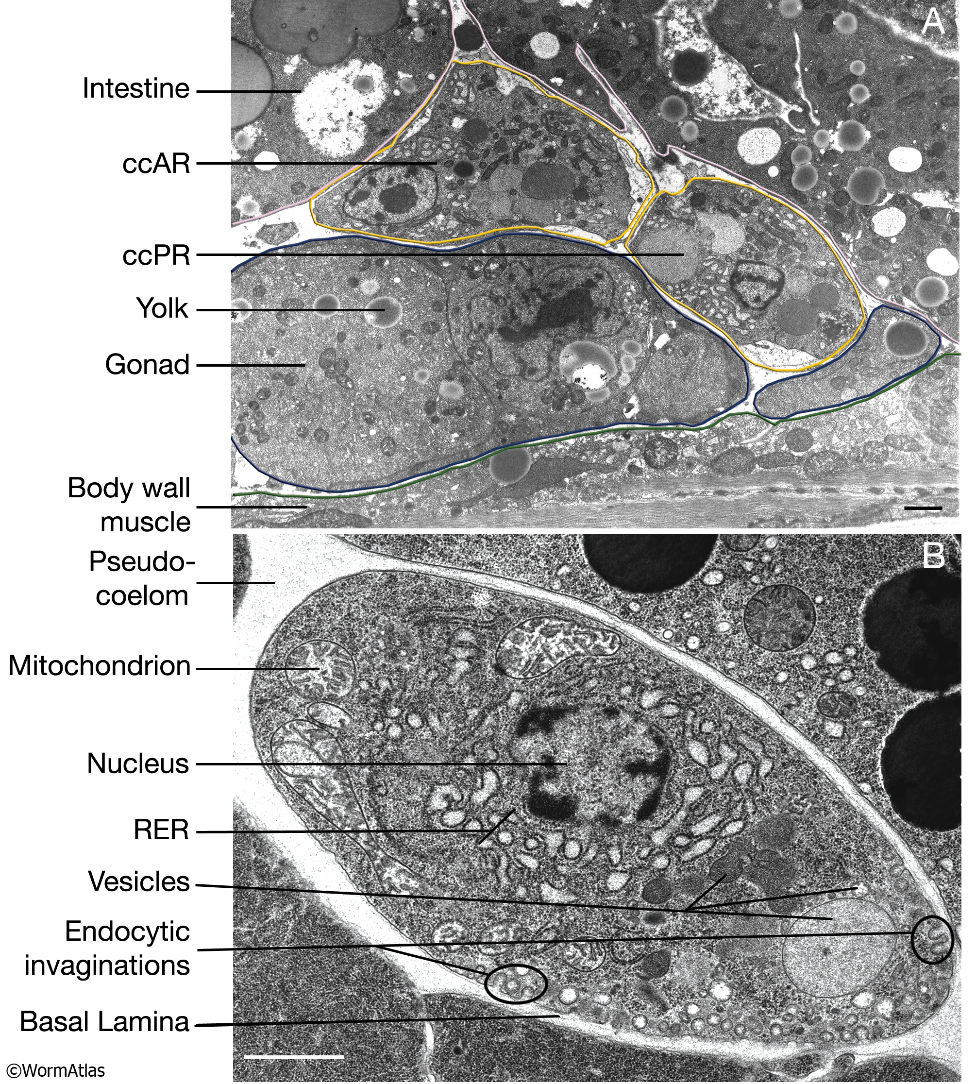
CcFIG 5 Legend
Transmission electron microscopy DNA sequencing - Google Transmission electron microscopy (TEM) produces high magnification, high resolution images by passing a beam of electrons through a very thin sample. Whereas atomic resolution has been demonstrated...

Transmission electron micrograph of the isolates KS-1 (a and d), KS-2 ...
Electron Micrographs of Cell Organelles | Zoology This is the electron micrograph of Lysosome, and is characterized by following features. These are also called Suicide bags or Death bags of the cell (Fig. 13 &14): (1) They were discovered by de Duve (1954). (2) They are spherical or irregular membrane bound vesicles filled with digestive enzymes.
Electron Micrograph of Actin and Intermediate Filaments In Part of a ...
Instruments of Microscopy | Microbiology | | Course Hero The transmission electron microscope (TEM) and scanning electron microscope (SEM) are two common forms. Scanning probe microscopy produces images of even greater magnification by measuring feedback from sharp probes that interact with the specimen. ... Label each component of the brightfield microscope.

Transmission electron micrographs of general structure. (A ...
Solved Label the transmission electron micrograph of the | Chegg.com Experts are tested by Chegg as specialists in their subject area. We review their content and use your feedback to keep the quality high. Answer The label is indicated from TOP to BOTTOM Ciliu …. View the full answer. Transcribed image text: Label the transmission electron micrograph of the cilium. Microvillus Axoneme Cilium Dynein arm.

Organelles | Biology for Majors I
IMG_2132.jpg - FIGURES Label this transmission electron micrograph ( 16 ... View Homework Help - IMG_2132.jpg from SCIENCE 101 at University High School, Tucson. FIGURES Label this transmission electron micrograph ( 16, 000 X ) of a relaxed sarcomere by placing the

4.4C: The Golgi Apparatus - Biology LibreTexts
anatomy 10.png - Label the transmission electron micrograph of the ... anatomy 10.png - Label the transmission electron micrograph of the. anatomy 10.png - Label the transmission electron micrograph... School Utah Valley University; Course Title ZOOL 1090; Uploaded By emileeroylance19. Pages 1 Ratings 67% (3) 2 out of 3 people found this document helpful;

Transmission electron micrographs showing the overall appearance of the ...
Electron microscopes - Cell structure - Edexcel - BBC Bitesize the scanning electron microscope (SEM) has a large depth of field. so can be used to examine the surface structure of specimens A human lymphocyte white blood cell as seen with a transmission ...
Lysosomes Dr.Jastrow's electron microscopic atlas
Solved Label this transmission electron micrograph of | Chegg.com Anatomy and Physiology questions and answers Label this transmission electron micrograph of relaxed sarcomeres by clicking and dragging the labels to the correct location Sarcamere 1 band (light) Z disc Mline Aband (dark) H zone
34 Label The Transmission Electron Micrograph Of The Nucleus - Labels 2021
Transmission electron microscopy characterization of ... - PubMed Transmission electron microscopy characterization of fluorescently labelled amyloid β 1-40 and α-synuclein aggregates BMC Biotechnol. 2011 Dec 19; 11:125. doi ... However, the use of these labels may interfere with the formation of larger-scale protein structures such as amyloid aggregates. Therefore, we investigate the effects of some ...

muscle tissues Dr.Jastrow's electron microscopic atlas
PDF Identifying Organelles from an Electron Micrograph The photograph shown below details chloroplast structure as viewed with a transmission electron microscope Courtesy of Dr. Julian Thorpe - EM & FACS Lab, Biological Sciences University Of Sussex A single Granum Chloroplast envelope visible as two membranes Stroma containing numerous small ribosomes Lamellae connecting different grana
Transmission electron micrograph of two viral DNA rings. Length two ...
Label The Transmission Electron Micrograph Of The Nucleus - Modern ... Answer to label the transmission electron micrograph of the nucleus. Labels are a means of identifying a product or container through a piece of fabric paper metal or plastic film onto which information about them is. The nucleus and nucleolus section 43 mitochondria section 410 and golgi apparatus section 47 can be seen.

PPT - Chapter 7 Notes PowerPoint Presentation, free download - ID:2252368
Electron Micrographs** Electron Micrographs**. Below is a collection of electron micrographs with labelled subcellular structures that you should be able to identify. Also, be sure to observe any electron micrographs which are made available in the laboratory by the instructor. You should concentrate on the similarities in form that permit identification of the ...

The Cell Nucleus Organization | Celebrate Cytochemistry | Gwen V ...
Transmission electron microscopy of specimens and processes in liquids ... Transmission electron microscopy is a powerful technique for the analysis of solid samples, but it can also be used to image in liquid environments, gaining a unique view of processes and structures in liquids. Here, we describe recent developments in electron microscopy of liquids and discuss applications in several areas. ... The QD labels ...

Structure and function of mitochondrial membrane protein complexes ...
Transmission electron microscopy - Wikipedia Transmission electron microscopy (TEM) is a microscopy technique in which a beam of electrons is transmitted through a specimen to form an image. The specimen is most often an ultrathin section less than 100 nm thick or a suspension on a grid. An image is formed from the interaction of the electrons with the sample as the beam is transmitted through the specimen.
Post a Comment for "38 label the transmission electron micrograph"