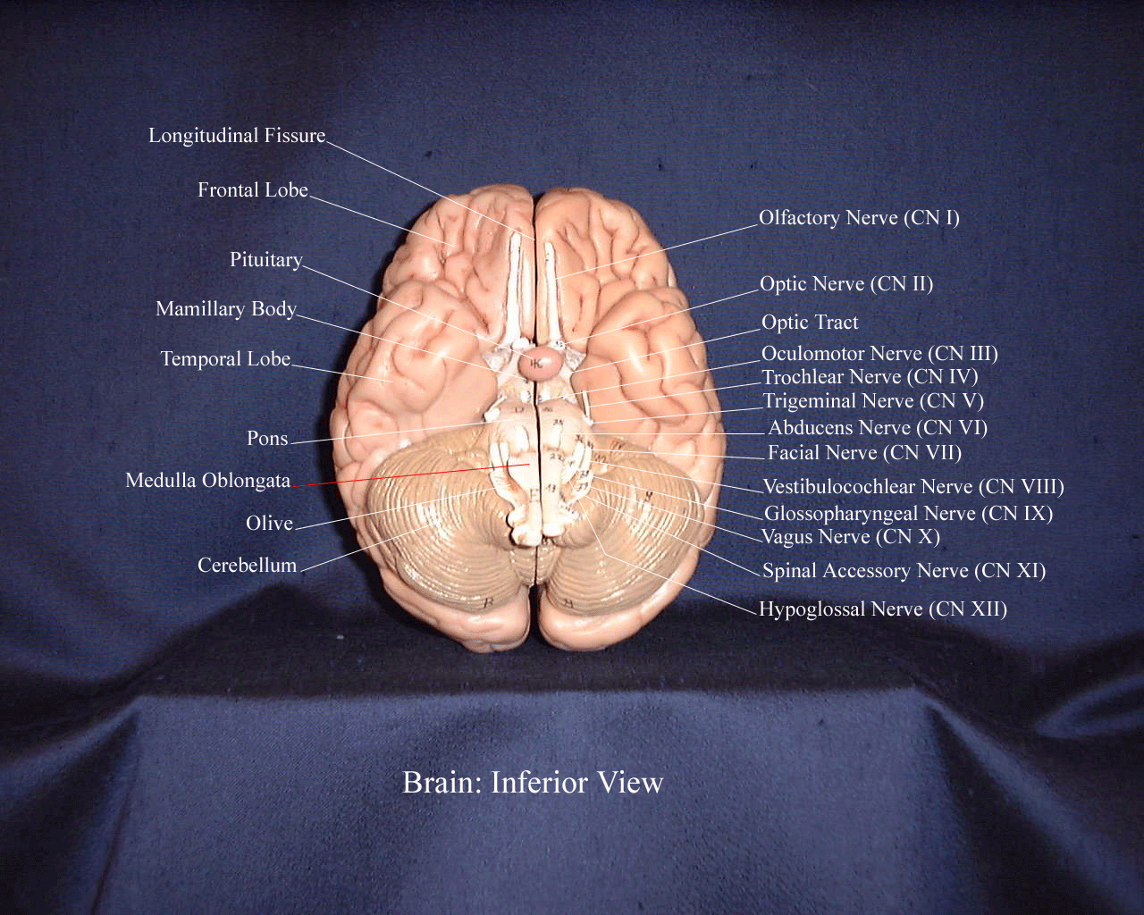
43 label the view of the inferior surface of the brain
Ch. 2 Surface of the Brain II Flashcards | Quizlet Label the parieto-occipital sulcus on the medial view. greater Label the prominent sulcus that forms the posterior boundary of the parietal lobe on the medial surface. The calcarine sulcus is the other prominent landmark on the medial surface of the occipital lobe. Label the horizontally oriented calcarine sulcus on the diagram. Directions, Reference Planes, & Views of the Brain - GetBodySmart The adult brain is often sectioned (cut) and viewed from different directions and angles. 1 2 Each point of view provides an altered perspective of the brain that changes the appearance of the major divisions, landmarks, and structures. Anatomical directions 1 2 3 4
Neuro Workbook Review ch 1-2 Flashcards - Quizlet This is a view of the ____al surface of the right cerebral cortex. The primary visual area is not visible because it is almost entirely in the walls of the calcarine fissure , on the _____ surface of the _____ lobe. Label the three indicated areas with the general class of functions in which each is involved.

Label the view of the inferior surface of the brain
precuneus: a review of its functional anatomy and behavioural ... A drawing of the medial surface of the human brain; the precuneus and its traditional anatomical landmarks are ... straight, T-shaped or more complex with three branches. The subparietal sulcus constitutes the inferior margin of the precuneus and continues its course around the posterior part of the cingulum. ... Their results led them to label ... The Midline Sagittal Surface of the Brain - Neuroscience - NCBI Bookshelf The parieto-occipital sulcus, running from the superior to the inferior aspect of the hemisphere, separates the parietal and occipital lobes. The calcarine sulcus divides the medial surface of the occipital lobe, running at very nearly a right angle from the parieto-occipital sulcus and marking the location of the primary visual cortex. Ventricles of the Brain: Anatomy, Function, CSF Flow - EZmed We will discuss the ventricles of the brain along with their anatomy and function, as well as the flow of cerebrospinal fluid (CSF) through those structures. By definition, the ventricles are cavities located within the brain that contain cerebrospinal fluid. The ventricles function to produce, circulate, and reabsorb CSF throughout the brain ...
Label the view of the inferior surface of the brain. Brainstem: Definition, anatomy, parts, function - Kenhub The brainstem (brain stem) is the distal part of the brain that is made up of the midbrain, pons, and medulla oblongata.Each of the three components has its own unique structure and function. Together, they help to regulate breathing, heart rate, blood pressure, and several other important functions.All of these brainstem functions are enabled because of its unique anatomy; since the brainstem ... Label Lateral View Of The Brain Quiz - PurposeGames.com About this Quiz. This is an online quiz called Label Lateral View Of The Brain. There is a printable worksheet available for download here so you can take the quiz with pen and paper. Total Points. 0. Duke Neurosciences - Lab 1: Surface Anatomy of the Brain At the inferior margin of the frontal lobe is the medial aspect of an inferior gyrus called the gyrus rectus (see 'ventral view' below), and a small division of the anterior and inferior cingulate gyrus, called the subcallosal (subgenual) area , which is just below the genu ("knee") of the corpus callosum. Brain: Anatomy, Pictures, Functions, and Conditions The brainstem is an area located at the base of the brain that contains structures vital for involuntary functions such as the heartbeat and breathing. The brain stem is comprised of the midbrain, pons, and medulla. Midbrain . The midbrain is often considered the smallest region of the brain. It acts as a sort of relay station for auditory and ...
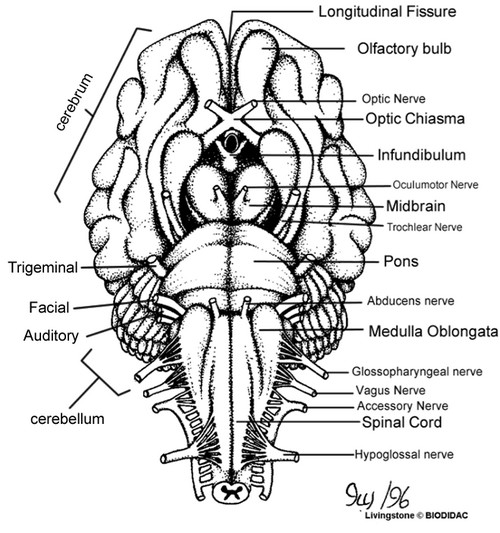
14.3 The Brain and Spinal Cord - Anatomy & Physiology The limbic cortex is the region of the cerebral cortex that is part of the limbic system, a collection of structures involved in emotion, memory, and behavior. Cerebral Cortex The cerebrum is covered by a continuous layer of gray matter that wraps around either side of the forebrain—the cerebral cortex. Lateral view of the brain: Anatomy and functions | Kenhub The pons is the middle part of the brainstem, located just inferior to the midbrain. It is connected with the cerebellum via the middle cerebellar peduncles. With its strategic relay position it is an important relay center between the cerebrum and the cerebellum. The trigeminal nerve (CN V) arises from the pons. How does the brain solve visual object recognition? - PMC Feb 09, 2012 · Specifically, the vast array of images caused by objects that should receive the same label (e.g. “car”, Fig. 1) results from the variability of the world and the observer: each object can be encountered at any location on the retina (position variability), at a range of distances (scale variability), at many angles relative to the observer ... Sheep Brain Dissection with Labeled Images - The Biology Corner Brain with Dura Mater Intact. Removal of the Dura Mater. 2. This image shows the ventral surface of the sheep's brain with most of the dura mater removed. The pituitary gland and the optic chiasma are still intact. (A = pituitary gland, B = optic chiasma, C = olfactory bulb) 3. On this image, the dura matter has been completely removed, you can ...
The Allen Mouse Brain Common Coordinate Framework: A 3D … May 14, 2020 · A surface view was then generated by considering only signal within 0.1 to 0.5 mm normalized cortical depths and projecting the largest maximum value to the surface. The virtual callosal labeling pattern further restricts boundaries of the higher visual areas, although to a lesser extent than in previous studies likely due to the density of ... Brain anatomy, Anatomy of the human brain | Mayfield Brain & Spine ... The brain is an amazing three-pound organ that controls all functions of the body, interprets information from the outside world, and embodies the essence of the mind and soul. Intelligence, creativity, emotion, and memory are a few of the many things governed by the brain. Protected within the skull, the brain is composed of the cerebrum ... Solved A Labeling 1· Label the view of the inferior surface - Chegg 100% (8 ratings) Answer: 11. Olive of Medulla …. View the full answer. Transcribed image text: A Labeling 1· Label the view of the inferior surface of the brain. 2. -16 8. 10. Sidman Neuroanatomy exam Flashcards | Quizlet This is a view of the _____al surface of the right cerebral cortex. The primary visual area is not visible because it is almost entirely in the walls of the calcarine fissure, on the _____ surface of the _____ lobe. Label the three indicated areas with the general class of functions in which each is involved.
Understanding Williams Syndrome: Chiari Malformation
Gas cylinder - Wikipedia A gas cylinder is a pressure vessel for storage and containment of gases at above atmospheric pressure.High-pressure gas cylinders are also called bottles.Inside the cylinder the stored contents may be in a state of compressed gas, vapor over liquid, supercritical fluid, or dissolved in a substrate material, depending on the physical characteristics of the contents.

Pin on Nursing
Picture of the Brain - WebMD The brain is one of the largest and most complex organs in the human body. It is made up of more than 100 billion nerves that communicate in trillions of connections called synapses. • The ...
InferiorBrainModel
Human Brain Diagrams and Detailed Information - Innerbody The cerebellum is a wrinkled, hemispherical region of the brain located posterior to the brainstem and inferior to the cerebrum. The outer layer of the cerebellum, known as the cerebellar cortex, is made of tightly folded gray matter that provides the processing power of the cerebellum.

Cerebrum: Inferior View Inferior Surface of Brain
The Dorsal and Ventral Surfaces of the Brain - Neuroscience - NCBI ... Dorsal view (A) and ventral view (B) of the human brain, indicating some of the major features visible from these perspectives. (C) The cerebral cortex has been removed in this dorsal view to reveal the underlying (After Rohen et al., (more...) The external features of the brain best seen on its ventralsurface are shown in

sheep brain dissection bi
Inferior view of the base of the skull: Anatomy | Kenhub The Jugular Foramen is a large j shaped foramen found in the recesses of the posterior cranial fossa, inferior to the petrous portion of the temporal bone. Posterior structures Inferior nuchal line (inferior view) On the outer surface of the occipital bone is the inferior nuchal line.

Sheep Brain Dissection Lab Companion
Anthropometry - Wikipedia Anthropometry (from Ancient Greek ἄνθρωπος (ánthrōpos) 'human', and μέτρον (métron) 'measure') refers to the measurement of the human individual. An early tool of physical anthropology, it has been used for identification, for the purposes of understanding human physical variation, in paleoanthropology and in various attempts to correlate physical with racial …
Cranial Nerves Coloring
File:Brain human normal inferior view with labels en.svg Original upload log []. File:Brain_human_normal_inferior_view.svg licensed with Cc-by-2.5 . 2009-10-13T16:18:05Z Beao 424x505 (209117 Bytes) Replaced right brain half with a clone of left brain half because they look excly the same in the picture.; 2007-09-23T15:14:17Z Ysangkok 424x505 (417241 Bytes) removing credits; 2007-03-03T17:30:01Z Ysangkok 424x505 (417718 Bytes) trying to make it work ...
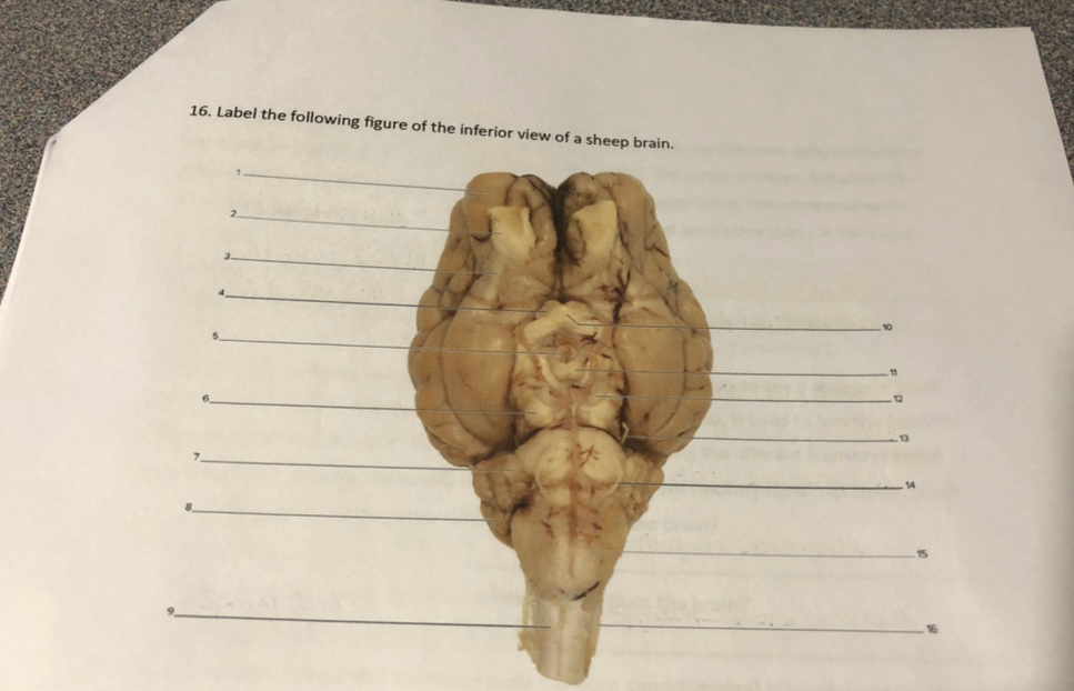
Solved: 1. Label The Following Figure Of The Inferior View... | Chegg.com
Duke Neurosciences - Lab 5: Forebrain Sectional Anatomy This most caudal ventricle in the adult brain lies between the dorsal surface of the pons and the large stalks of white matter (the cerebellar peduncles; "peduncle" means stalk) that connect the cerebellum to the brainstem. Now return to the forebrain and view the midsagittal plane (go to Sectional Anatomy > Photographic Atlas > Ventricles ...

Lateral View of the Brain Quiz - PurposeGames
WAYS TO VIEW THE BRAIN - Richards on the Brain Below the midbrain, caudal = inferior. Dorsal: pertaining to the back or upper surface of the body; opposite of "ventral." (NCIt) Structures found on the top of the human brain or on the top of some other structure within the brain. (Kolb, 39) Editor's note - from the Latin word for 'back' as in the location of a shark's fin.
Post a Comment for "43 label the view of the inferior surface of the brain"