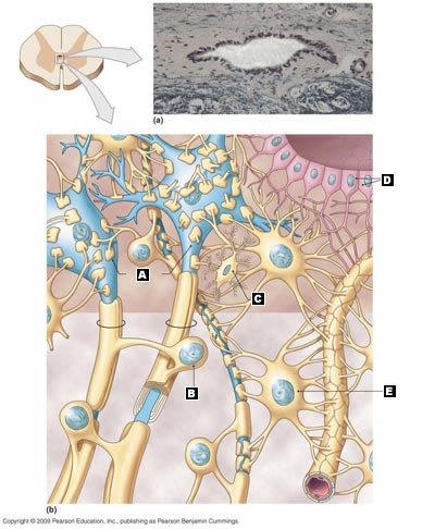
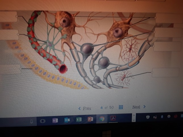
39 correctly label the following anatomical features of the neuroglia
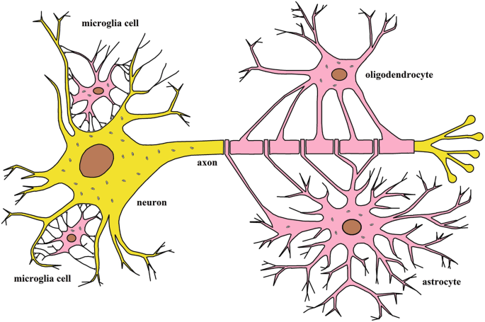
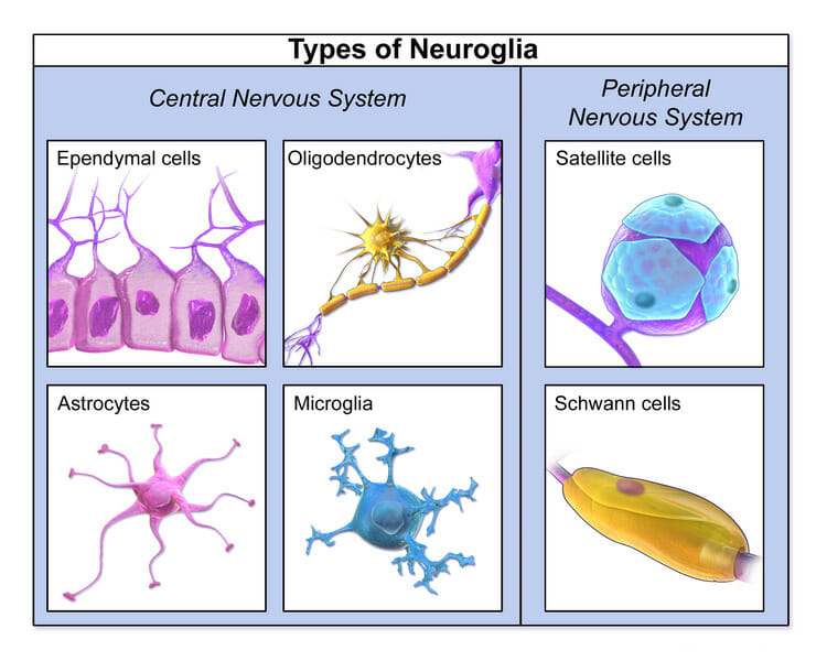
Answered: Describe the structures and functions… | bartleby Describe the structures and functions of the neurons and neuroglia of the cerebrum, the cerebellum, the diencephalon, and the brain stem. Describe the structures and functions of the Schwann Cells in both the central nervous system and the peripheral nervous system. What role does the Pituitary gland play as the control center of the brain? Difference Between Neurons And Neuroglia - An Overview Neuroglia in the central nervous system include astrocytes, oligodendrocytes, microglial cells, and ependymal cells. Schwann cells and satellite cells are the neuroglia in the peripheral nervous system. Also Read: Neurons Neurons are the structural and functional unit of nervous system. They help in transmitting the nerve impulse.
Neuroglia | Boundless Anatomy and Physiology | | Course Hero Neuroglia in the CNS include astrocytes, microglial cells, ependymal cells and oligodendrocytes. Neuroglia in the PNS include Schwann cells and satellite cells.Astrocytes support and brace the neurons and anchor them to their nutrient supply lines. They also play an important role in making exchanges between capillaries and neurons.
Correctly label the following anatomical features of the neuroglia
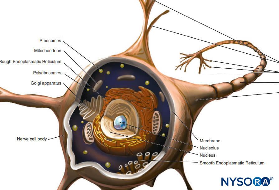
Nervous Tissue Glial Cells - ThoughtCo Neuroglia, also called glia or glial cells, are non-neuronal cells of the nervous system. They compose a rich support system that is essential to the operation of nervous tissue and the nervous system. Unlike neurons, glial cells do not have axons, dendrites, or conduct nerve impulses. A Labelled Diagram Of Neuron with Detailed Explanations They are found in the brain, spinal cord and the peripheral nerves. A neuron is also known as the nerve cell. The structure of a neuron varies with their shape and size and it mainly depends upon their functions and their location. Neurons are the structural and functional units of the nervous system. A group of neurons forms a nerve. Overview of neuron structure and function - Khan Academy Anatomy of a neuron. Neurons, like other cells, have a cell body (called the soma ). The nucleus of the neuron is found in the soma. Neurons need to produce a lot of proteins, and most neuronal proteins are synthesized in the soma as well. Various processes (appendages or protrusions) extend from the cell body.
Correctly label the following anatomical features of the neuroglia. Neuron Anatomy, Nerve Impulses, and Classifications - ThoughtCo A neuron consists of two major parts: a cell body and nerve processes. Cell Body Neurons contain the same cellular components as other body cells. The central cell body is the process part of a neuron and contains the neuron's nucleus, associated cytoplasm, organelles, and other cell structures. Labeled Neuron Diagram | Science Trends Neurons are a type of cell and are the fundamental constituents of the nervous system and brain. Neurons take in stimuli and convert them to electrical and chemical signals that are sent to our brain. There are 3 major kinds of neurons in the spinal cord: sensory, motor, and interneurons. Neurons communicate vie electrical signals produced by ... Neuron Diagram & Types - Ask A Biologist Nerve cells are also some of the longest cells in your body. There are nerve cells as long as a meter. They stretch from your hips all the way down to your toes! This is very uncommon for cells, which are usually very short. Most cells are 20 micrometers in diameter, which is just a fraction of the width of a hair. Neuron Anatomy Solved Neurons and neuroglia Correctly label the following - Chegg Question: Neurons and neuroglia Correctly label the following anatomical features of nervous tissue in the brain and spinal cord. Microglia Cell body Neuron Capillary Dendrite Myelin sheath Astrocyte Oligodendrocyte Axon Nucleus This problem has been solved! See the answer Show transcribed image text Expert Answer 100% (8 ratings)
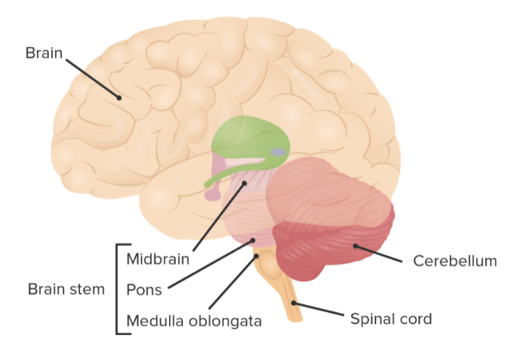
Nervous System - Label the Brain - TheInspiredInstructor.com This brain part controls thinking. This brain part controls balance, movement, and coordination. This brain part controls involuntary actions such as breathing, heartbeats, and digestion. This part of the nervous system moves messages between the brain and the body. This part of the cerebrum interprets and sorts information from the senses. BIO215 Week 2 - Wesleyan College 3) List and describe the characteristic features and rules for naming connective tissue proper . 4) Recognize in microscope slides, correctly label, and describe the major properties and locations of the major types of connective tissue proper. correctly label the following anatomical features of a nerve Solved ... correctly label the following anatomical features of a nerve Solved: correctly label the following anatomical features. Kapsels 2020 halflang bruin kapsel halflang 2020 Kapsels halflange nieuwste haarkleuren Fijn halflang ReaderUi ReaderUi
Neuroglia - Definition, Functions, Types | Solved Question The Neuroglia are a group of supportive cells for the neurons. Further, they maintain the myelin sheath, provide nutrient support. Moreover, they also retain homeostasis. It is within the CNS and PNS. Neuroglia Functions It offers essential nutrients. It includes oxygen to neurons. Next, it also helps create the myelin sheath. Anatomy QA- Neuroglial cells: structure and function Following are the functions of neuroglial cells: Provide structural support to neurons. They Form myelin sheath. Participate in formation of blood - brain barrier. Phagoctytosis. Produce cerebrospinal fluid. Name the various neuroglial cells. A. Neuroglia in Central nervous system (CNS) 1. Asrtocytes They are the largest glial cells. Nervous system: Structure, function and diagram | Kenhub The nervous system is a network of neurons whose main feature is to generate, modulate and transmit information between all the different parts of the human body. This property enables many important functions of the nervous system, such as regulation of vital body functions ( heartbeat, breathing, digestion), sensation and body movements. Answered: 5 | bartleby Related Anatomy and Physiology Q&A. Find answers to questions asked by students like you. Show more Q&Aadd. question_answer. Q: Correctly label the following veins of the thorax. Hemiazygos v. Internal jugular v. ... The neurons and the supporting cells known as neuroglia form the nervous tissue. The neuralgia cells...
COGNITIVE NEUROSCIENCE THE BIOLOGY OF THE MIND Fourth … Academia.edu uses cookies to personalize content, tailor ads and improve the user experience. By using our site, you agree to our collection of information through the use of cookies.
Ch 12 - Nervous System EXAM *** McGraw Flashcards - Quizlet Correctly label the following anatomical features of the neuroglia. presynaptic terminals. Synaptic vesicles contain neurotransmitters and are present in the Can conduct an impulse Which of the following is NOT characteristic of neuroglia? from node to node on a myelinated axon. Action potentials are conducted more rapidly when transmission is
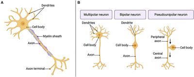
Chapter 12 QS Anatomy (Nervous System) Flashcards - Quizlet Match these definitions to the correct term. 1. Many dendrites and a single axon (Click to select) 2. One dendrite and one axon (Click to select) 3. One process with two branches; one extending to the CNS, one extending to the periphery (Click to select) 1. Many dendrites and a single axon (Multipolar neuron) 2.
PDF Gross Anatomy of the Brain and Cranial Nerves - Pearson nerves (PNS) because of their close anatomical relationship. The Human Brain During embryonic development of all vertebrates, the CNS first makes its appearance as a simple tubelike structure, the neural tube, that extends down the dorsal median plane. By the fourth week, the human brain begins to form as an expan-
THE NERVOUS SYSTEM - Lake County Schools / Overview - AnyFlip Key Choices B. Neuroglia 1. Support, insulate, and protect cells A. Neurons 2. Demonstrate irritability and conductivity, and thus transmit electrical messages from one area of the body to another area 3. Release neurotransmitters 4. Are amitotic S. Able to divide; therefore are responsible for most brain neoplasms Chapter 7 The Nervous System 133
Anatomy Worksheet 1.0... - Course Hero the microscopic structure of skeletal muscle tissue using chapter 10 of your text and a skeletal muscle tissue slide, complete the following. draw and label the following structures: sarcomere,sarcolemma,z disc,a band, i band, m line, actin andmyosin (many features in the slide will be too small to see clearly/at all obtain a smooth muscle …
NEURON STRUCTURE AND CLASSIFICATION - Brigham Young University-Idaho Structural classification of neurons. 1) Bipolar; 2) Multipolar and 3) Unipolar. Bipolar neurons have only two processes that extend in opposite directions from the cell body. One process is called a dendrite, and another process is called the axon. Although rare, these are found in the retina of the eye and the olfactory system.
Nervous System Anatomy and Physiology - Nurseslabs Supporting cells in the CNS are "lumped together" as neuroglia, literally mean "nerve glue". Neuroglia. Neuroglia include many types of cells that generally support, insulate, and protect the delicate neurons; in addition, each of the different types of neuroglia, also simply called either glia or glial cells,has special functions. Astrocytes.
Solved Correctly label the following anatomical features of - Chegg Question: Correctly label the following anatomical features of the neuroglia. Axon terminal Neurons Oligodendrocyte Astrocyte Microglia Myelinated axon Capillary Schwann cell Ependymal cell Satellite cell ag Poble o 2000 Reset Zoom < Prev 13 of 55 Next > This problem has been solved! See the answer Show transcribed image text Expert Answer
Correctly Label The Following Anatomical Features Of A Neuron First, the axon is a long, thin tube. Some of these branches are connected by a synapses called dendrites. In humans, the axon is over a meter long. Another important feature of a neuron is its synapse, a region of the plasma membrane that contains a single, continuous sarcomere. The nucleus of a neuron is large and round.
The Neuron - BrainFacts Cells within the nervous system, called neurons, communicate with each other in unique ways. The neuron is the basic working unit of the brain, a specialized cell designed to transmit information to other nerve cells, muscle, or gland cells.
Overview of neuron structure and function - Khan Academy Anatomy of a neuron. Neurons, like other cells, have a cell body (called the soma ). The nucleus of the neuron is found in the soma. Neurons need to produce a lot of proteins, and most neuronal proteins are synthesized in the soma as well. Various processes (appendages or protrusions) extend from the cell body.
A Labelled Diagram Of Neuron with Detailed Explanations They are found in the brain, spinal cord and the peripheral nerves. A neuron is also known as the nerve cell. The structure of a neuron varies with their shape and size and it mainly depends upon their functions and their location. Neurons are the structural and functional units of the nervous system. A group of neurons forms a nerve.
Nervous Tissue Glial Cells - ThoughtCo Neuroglia, also called glia or glial cells, are non-neuronal cells of the nervous system. They compose a rich support system that is essential to the operation of nervous tissue and the nervous system. Unlike neurons, glial cells do not have axons, dendrites, or conduct nerve impulses.


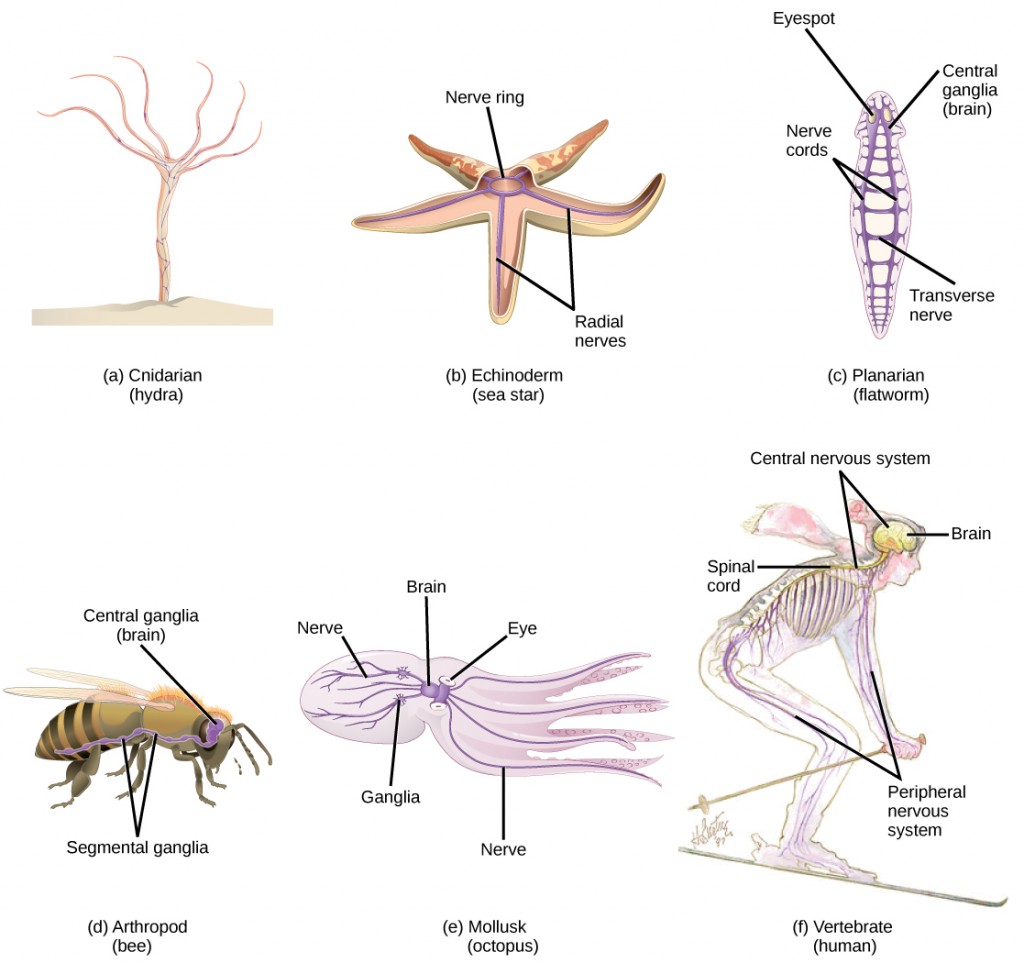



:watermark(/images/watermark_only_sm.png,0,0,0):watermark(/images/logo_url_sm.png,-10,-10,0):format(jpeg)/images/anatomy_term/parasympathetic-nervous-system/NW2chCFVkZmbGDJc7RjXGg_Parasympathetic_nervous_system.jpeg)






Post a Comment for "39 correctly label the following anatomical features of the neuroglia"