38 microscope labeled
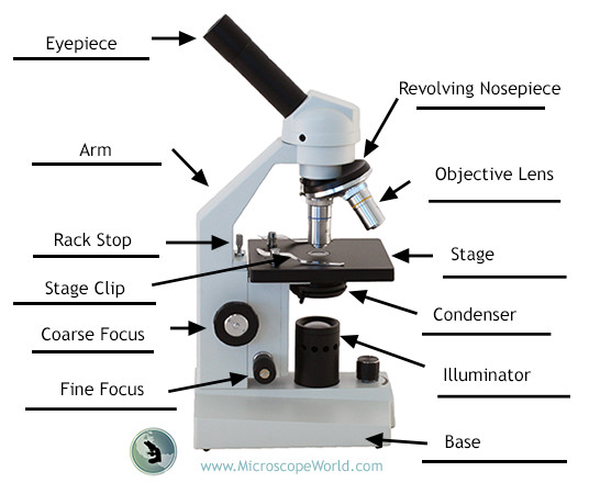
Sperm Under Microscope with Labeled Diagram - AnatomyLearner Spermatogonia under the light microscope In the germinal epithelium of a seminiferous tubule, you will find spermatogonia (stem cells) at its base. Again, the other spermatogenic cells are arranged in the order of the development process. The spermatogonia of the seminiferous tubules are immature cells that undergo several mitotic divisions. Parts of the Microscope with Labeling (also Free Printouts) Parts of the Microscope with Labeling (also Free Printouts) By Editorial Team March 7, 2022 A microscope is one of the invaluable tools in the laboratory setting. It is used to observe things that cannot be seen by the naked eye. Table of Contents 1. Eyepiece 2. Body tube/Head 3. Turret/Nose piece 4. Objective lenses 5. Knobs (fine and coarse) 6.
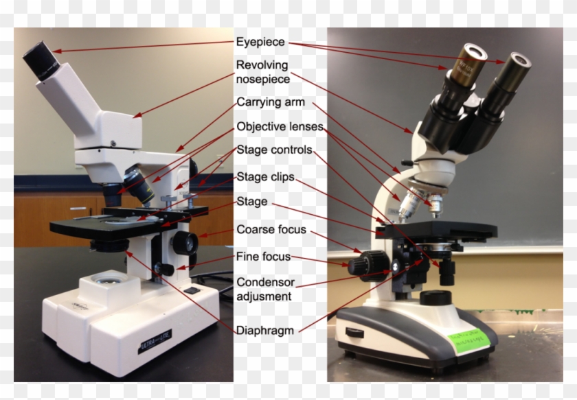
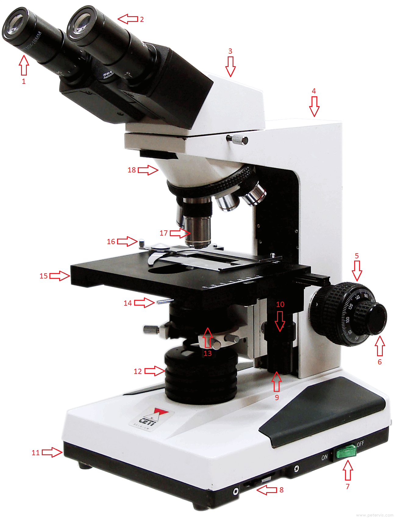
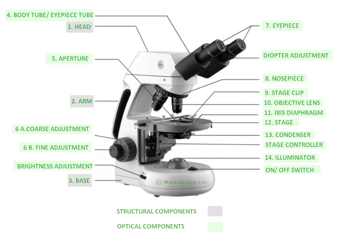
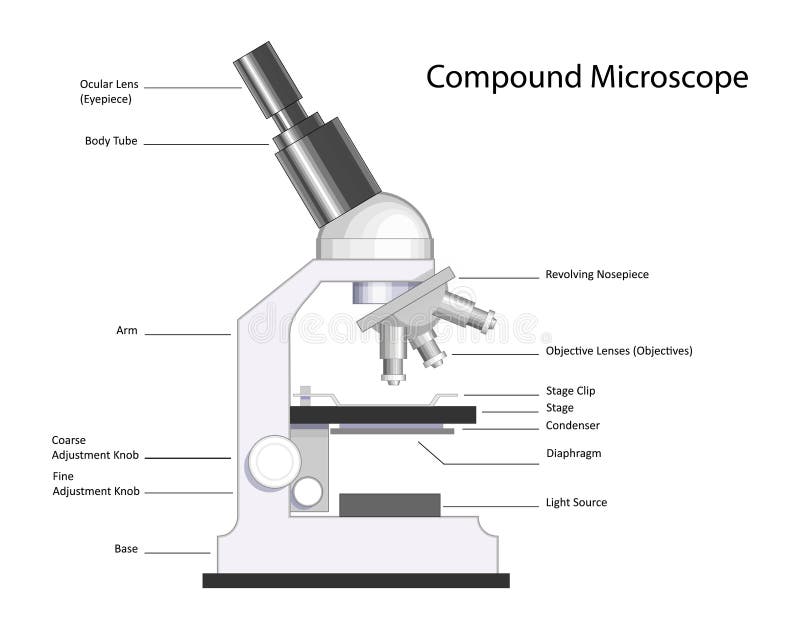
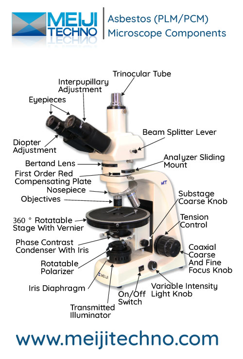
Compound Microscope Parts - Labeled Diagram and their Functions Labeled diagram of a compound microscope Major structural parts of a compound microscope There are three major structural parts of a compound microscope. The head includes the upper part of the microscope, which houses the most critical optical components, and the eyepiece tube of the microscope.

Microscope labeled
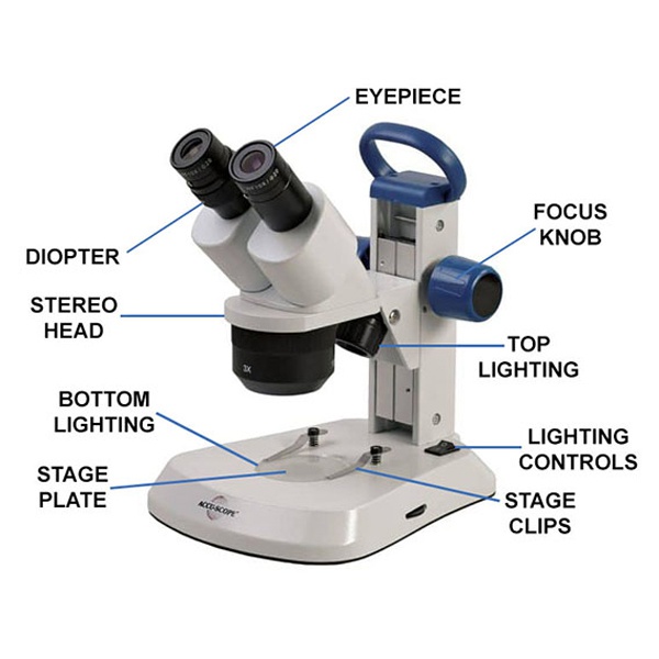
Parts of Stereo Microscope (Dissecting microscope) - labeled diagram ... Major microscope brands (Zeiss, Olympus, Nikon, Amscope, Omano, Leica …) all produce stereomicroscopes. Photo source: AOMEKIE, Bresser, Swift The name "stereo" comes from the term "stereoscopic," meaning, viewing by two different angles to create an impression of depth and solidity. Microscope Parts and Functions Most specimens are mounted on slides, flat rectangles of thin glass. The specimen is placed on the glass and a cover slip is placed over the specimen. This allows the slide to be easily inserted or removed from the microscope. It also allows the specimen to be labeled, transported, and stored without damage. Microscope Types (with labeled diagrams) and Functions Simple microscope labeled diagram Simple microscope functions It is used in industrial applications like: Watchmakers to assemble watches Cloth industry to count the number of threads or fibers in a cloth Jewelers to examine the finer parts of jewelry Miniature artists to examine and build their work Also used to inspect finer details on products
Microscope labeled. Microscope With Labeled Parts and Functions - 24 Hours Of Biology Microscope is a revolutionized scientific instrument which is used in research laboratories to examine the small objects that are not clearly visible and can't be seen by the naked eye. They are derived from Ancient Greek words "mikrós skopeîn" as mikrós means "small" and skopeîn mean "to look" or "see". Microscope Parts, Function, & Labeled Diagram - slidingmotion Microscope Parts Labeled Diagram The principle of the Microscope gives you an exact reason to use it. It works on the 3 principles. Magnification Resolving Power Numerical Aperture. Parts of Microscope Head Base Arm Eyepiece Lens Eyepiece Tube Objective Lenses Nose Piece Adjustment Knobs Stage Aperture Microscopic Illuminator Condenser Lens (PDF) Light and electron microscopic study of fetal lung following ... examined with a Leitz-Dialux-20 microscope. Black and . ... ribosomal subunits and polyribosomes along with a three-fold increase in the incorporation of labeled amino acids indicates an induction ... Microscope, Microscope Parts, Labeled Diagram, and Functions Microscope, Microscope Parts, Labeled Diagram, and Functions What is Microscope? A microscope is a laboratory instrument used to examine objects that are too small to be seen by the naked eye. It is derived from Ancient Greek words and composed of mikrós, "small" and skopeîn,"to look" or "see".
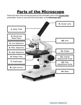
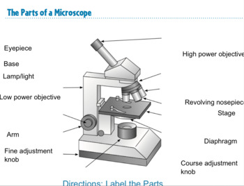
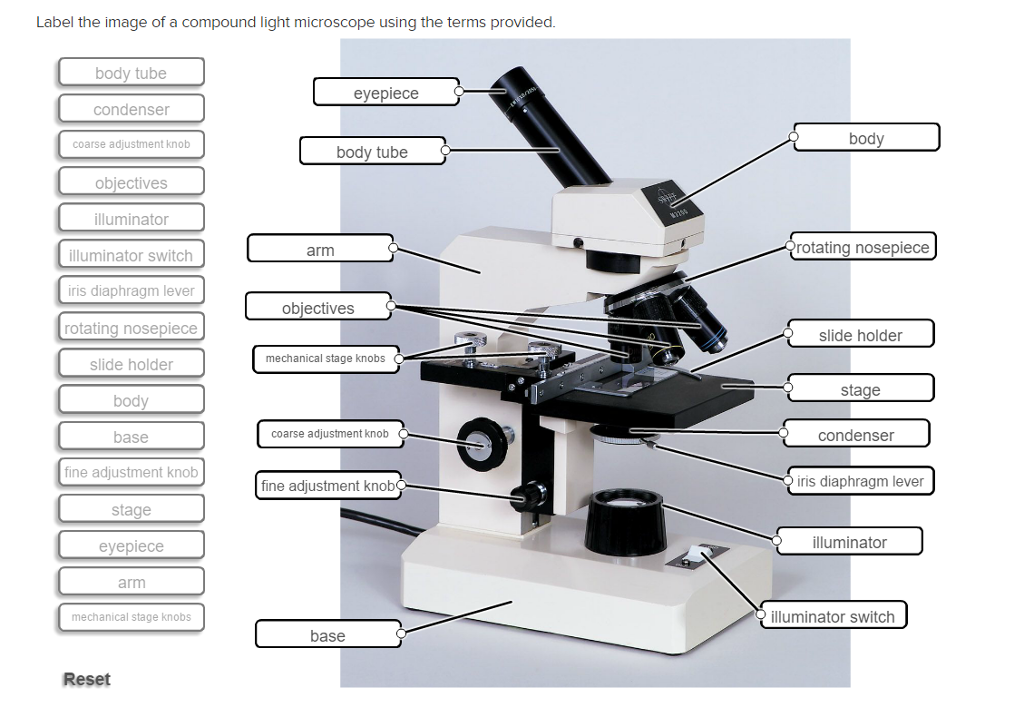
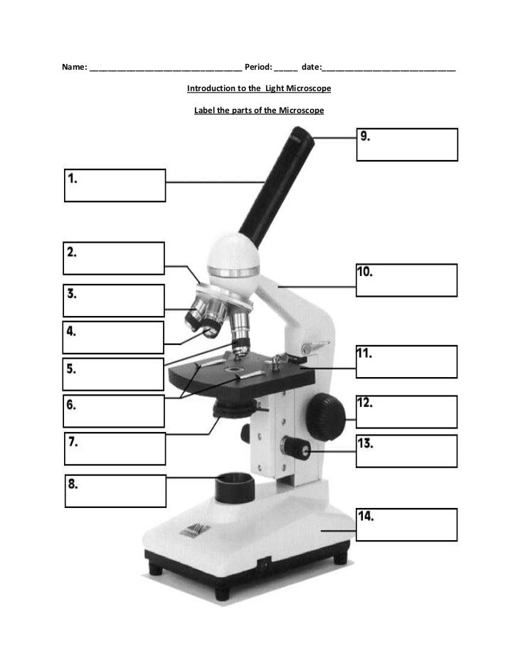
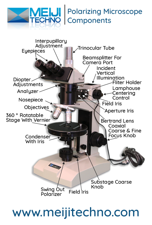
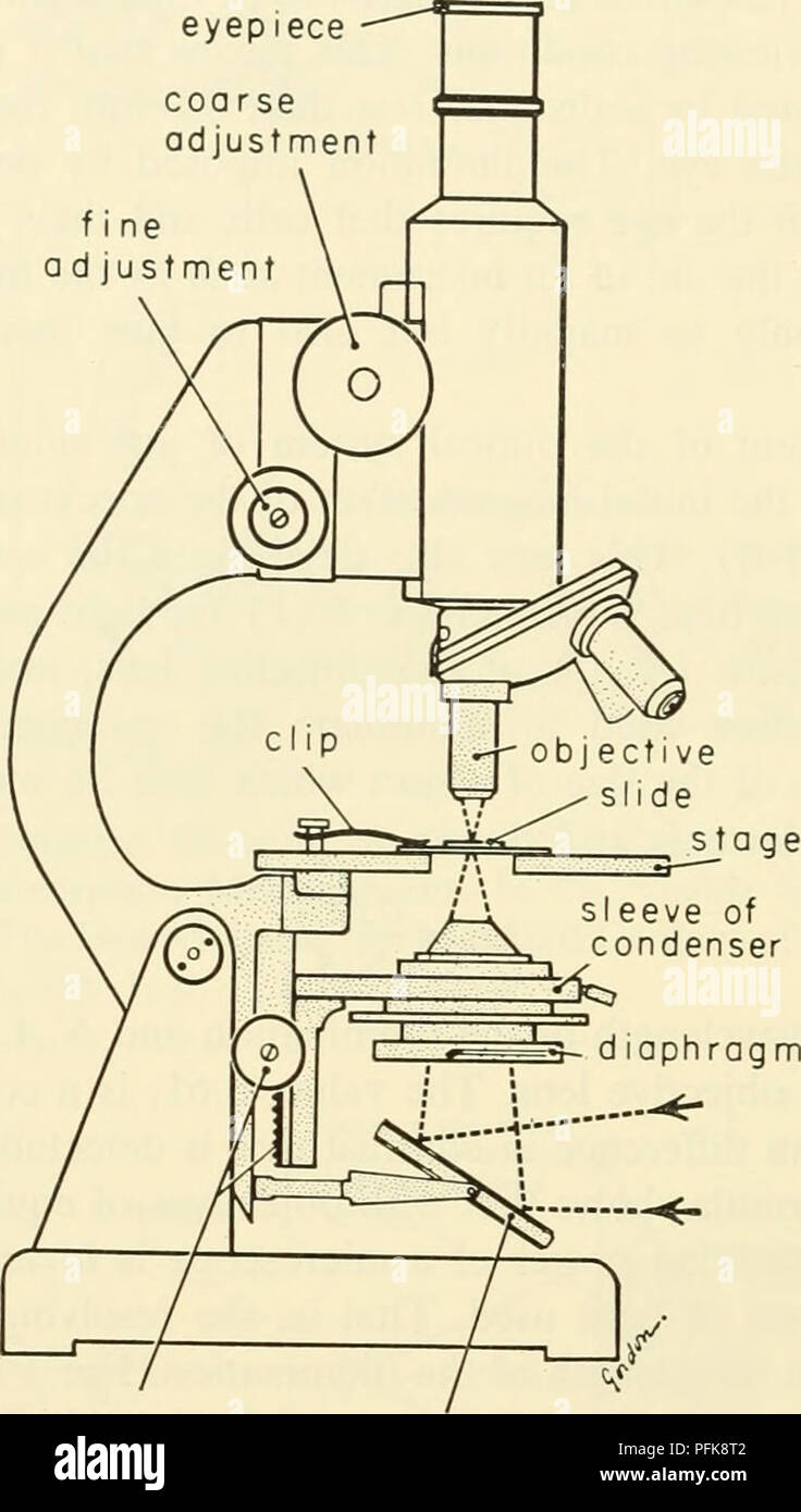
Atherosclerosis diagnostic imaging by optical spectroscopy ... - DeepDyve Atherosclerosis is traditionally viewed as a disease of uncontrolled plaque growth leading to arterial occlusion. More recently, however, occlusion of the arterial lumen is being viewed as an acute event triggered by plaque rupture and thrombosis. An atheromatous plaque becomes vulnerable to sudden activation and/or rupture when a constellation of processes are activated by various trigger ... Microscope Labeling - The Biology Corner The labeling worksheet could be used as a quiz or as part of direct instruction where students label the microscope as you go over what each part is used for. The google slides shown below have the same microscope image with the labels for students to copy. Compound Microscope Parts, Functions, and Labeled Diagram Common compound microscope parts include: Eyepiece (ocular lens) with or without Pointer: The part that is looked through at the top of the compound microscope. Eyepieces typically have a magnification between 5x & 30x. Monocular or Binocular Head: Structural support that holds & connects the eyepieces to the objective lenses. A Study of the Microscope and its Functions With a Labeled Diagram ... To better understand the structure and function of a microscope, we need to take a look at the labeled microscope diagrams of the compound and electron microscope. These diagrams clearly explain the functioning of the microscopes along with their respective parts. Man's curiosity has led to great inventions. The microscope is one of them.
Nonlinear optical microscopy in decoding arterial diseases - ResearchGate The capability of label-free microscopic imaging enables disease impact to be studied directly on the bulk artery tissue, thus minimally perturbing the sample. ... timodal CARS microscope with fs ... Motorized Rotary Microtomes Suppliers @ MedicRegister.com Fisher Scientific International, Inc. | Address: Liberty Lane, Hampton, New Hampshire 03842, USA | Send Inquiry | Phone: +1-(603)-926-5911 FDA Registration: 2431530 Annual Revenues: USD 50-100 Million Employee Count: ~3300 Products: Slide Staining, General Medical Supplies, General Examination Supplies, Manual Slide Staining System, Microscope Slide Staining Kit ... Label the microscope — Science Learning Hub All microscopes share features in common. In this interactive, you can label the different parts of a microscope. Use this with the Microscope parts activity to help students identify and label the main parts of a microscope and then describe their functions. Drag and drop the text labels onto the microscope diagram. Parts of a microscope with functions and labeled diagram - Microbe Notes Microscopes are instruments that are used in science laboratories to visualize very minute objects such as cells, and microorganisms, giving a contrasting image that is magnified. Microscopes are made up of lenses for magnification, each with its own magnification powers.
Microscope Labeled Pictures, Images and Stock Photos Microscope Labeled Pictures, Images and Stock Photos View microscope labeled videos Browse 49 microscope labeled stock photos and images available, or start a new search to explore more stock photos and images. Newest results Fluorescent Imaging immunofluorescence of cancer cells growing... Microscope diagram vector illustration.
Simple Microscope - Diagram (Parts labelled), Principle, Formula and Uses A simple microscope consists of Optical parts Mechanical parts Labeled Diagram of simple microscope parts Optical parts The optical parts of a simple microscope include Lens Mirror Eyepiece Lens A simple microscope uses biconvex lens to magnify the image of a specimen under focus.
Microscope Types (with labeled diagrams) and Functions Simple microscope labeled diagram Simple microscope functions It is used in industrial applications like: Watchmakers to assemble watches Cloth industry to count the number of threads or fibers in a cloth Jewelers to examine the finer parts of jewelry Miniature artists to examine and build their work Also used to inspect finer details on products
Microscope Parts and Functions Most specimens are mounted on slides, flat rectangles of thin glass. The specimen is placed on the glass and a cover slip is placed over the specimen. This allows the slide to be easily inserted or removed from the microscope. It also allows the specimen to be labeled, transported, and stored without damage.
Parts of Stereo Microscope (Dissecting microscope) - labeled diagram ... Major microscope brands (Zeiss, Olympus, Nikon, Amscope, Omano, Leica …) all produce stereomicroscopes. Photo source: AOMEKIE, Bresser, Swift The name "stereo" comes from the term "stereoscopic," meaning, viewing by two different angles to create an impression of depth and solidity.















Post a Comment for "38 microscope labeled"