39 how to label nmr spectra
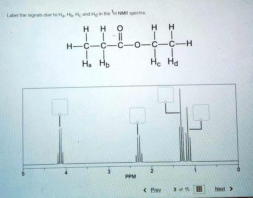
MetaboAnalyst Urinary metabolite concentrations from 77 cancer patients measured by 1H NMR. Phenotype: N - cachexic; Y - control Solved 4H 6H 4H 3 ppm PPM 5. Use your 'H - NMR spectrum to - Chegg Using your IR and NMR data. Question: 4H 6H 4H 3 ppm PPM 5. Use your 'H - NMR spectrum to determine how many different types of hydrogens are present in your unknown molecule. (I point) 6. On your NMR spectra, label the integration and splitting pattern type (singlet, doublet, triplet, quartet, multiplet ete) for each signal of your 'H-NMR ...
EPR - Interpretation - Chemistry LibreTexts Web16-04-2022 · Proportionality factor (g-factor) As a result of the Zeeman Effect, the state energy difference of an electron with s=1/2 in magnetic field is \[ \Delta E = gβB \label{1}\] where β is the constant, Bohr magneton. Since the energy absorbed by the electron should be exactly the same with the state energy difference ΔE, ΔE=hv ( h is Planck’s constant), …
How to label nmr spectra
Starting guide to Mnova 1D-NMR spectrum - Mestrelab Resources To do this go to 'NMR/Processing' and then apply 'Auto baseline correction'. This spectrum is now ready for analysis so go now to 'NMR/Analysis' and select 'Reference' to make sure that this peak is set to the suitable chemical shift. Now, click 'Auto Multiplet Analysis' button to carry out Peak picking, Integration and ... Introduction to CREST — xtb doc 2021 documentation - Read the … WebOther standalone functionalities that are included in CREST are parallel optimization and screening functions for GFNn–xTB, the function to sort (e.g. for NMR equivalencies) externally created ensembles, and some automated procedures for the protonation, deprotonation and tautomerization of structures. MetaboAnalyst Group Label. Description: Data 1 ... Common name: Discrete: Urinary metabolite concentrations from 77 cancer patients measured by 1H NMR. ... You can still process ...
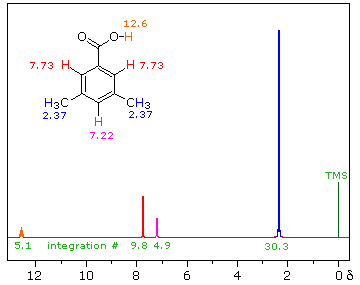
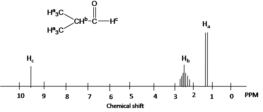
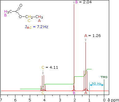
How to label nmr spectra. How To Analyze The Peaks Of H-NMR Spectroscopy - YouTube Presents: H-NMR How To Analyze PeaksAre you struggling with organic chemistry? Download my free ebook "10 Secrets To Aci... 13C Carbon NMR Spectroscopy - Chemistry Steps In general, when you start analyzing a 13 C NMR, split the spectrum in two parts by drawing a line at 100 ppm; below this value you have the saturated functional groups, and beyond that is the unstructured region. Saturated carbon atoms connected to electronegative heteroatoms give signal from 30-90 ppm. The Basics of Interpreting a Proton (1H) NMR Spectrum - ACD/Labs For a 1 H-NMR spectrum to be consistent with a chemical structure, the following must all be true: All chemical shifts are in the appropriate range for the assigned protons' local magnetic environment All peaks exhibit the appropriate multiplicity All peaks exhibit an appropriate integral value 6.6 ¹H NMR Spectra and Interpretation (Part I) WebAcetaldehyde: The three H a protons in the methyl group are chemical equivalent, and they all bonded to an sp 3-hybridized carbon; but they are different from the H b proton that is bonded to an sp 2 –hybridized carbonyl carbon. There are two total signals in the 1 H NMR spectrum.; 1,4-dimethylbenzene: all four aromatic protons are chemically equivalent …
NMR Spectrum of Aspirin | Thermo Fisher Scientific - US NMR Spectrum of Aspirin Aspirin (C 9 H 8 O 4 ), acetylsalicylic acid, is a salicylate drug used to treat a variety of conditions, including headaches, fever and inflammation. It is also used in low doses in preventive treatment against heart attacks, strokes and blood clots. Collaborative Computational Project for NMR - CCPN WebThe programme lets you view, inspect, peak pick, label and assign one or many NMR spectra, either 1D or nD up to 8 dimensions. See the features. AnalysisScreen. Designed for NMR-based small-molecule screening, AnalysisScreen lets you view and analyse large 1D data sets, find hits and create compound mixtures. NMR Sample Preparation | Chemical Instrumentation Facility ... Label your sample. Permanent marker can be used to label the sample to ensure you do not mix up your NMR tube with another NMR user, or it can be retrieved at a later time. If you use a sticker or tape to label the tube, the sticker must be flush with the tube so it does not interfere with insert, eject or spinning inside the magnet. PDF Manual - Practical NMR 39 Experiment 4 Practical NMR: Collecting and Interpreting Your Own Spectra Reading: Handbook for Organic Chemistry Lab, chapters on NMR Spectroscopy (Chapter 18) and Identification of Compounds (Chapter 20). Organic Chemistry, Marc Loudon, 6th ed., pp. 611-653 (13.1-13.7); pp. 662-667 (13.10-13.11). Previously, you learned how to interpret the 1H NMR spectra of pure compounds.
Isotopic Labeling for NMR Spectroscopy of Biological Solids The simplest and most cost-effective biosynthetic labeling method for protein solid-state NMR is to uniformly label all carbon and nitrogen atoms with 13 C and 15 N. In this way, a single protein sample can in principle provide all the structural constraints - dihedral angles and distances - about the protein. MetaboAnalyst WebGroup Label: Categorical (Classification) Continuous ... Description: Dataset: Common name: Discrete: Urinary metabolite concentrations from 77 cancer patients measured by 1H NMR. Phenotype: N - cachexic; Y - control ... Sorry. User login module is being upgraded. You can still process your spectra without login. We will come back ... Simulate and predict NMR spectra Predict 1D 13 C NMR spectra. Predict COSY spectra. Predict HSQC / HMBC spectra. Simulated second order effect in 1 H NMR spectra. Recreate (resurect) 1D NMR spectra from experimental in-line assignment. 1 H NMR prediction was possible thanks to the tool of the FCT-Universidade NOVA de Lisboa developped by Yuri Binev and Joao Aires-de-Sousa. Y. MetaboAnalyst WebThe class label must be in the second row. The requirements for class ... The screenshot below shows a subset of binned NMR spectra data (bin width 0.04 ppm). In this table, the samples from controls (e.g. Contr_1) are paired with the samples from the subjects in disease (Disease_1) based on some criteria (i.e. age, weight ...
Chapter 1: NMR Coupling Constants - orgchemboulder.com Chapter 1: NMR Coupling Constants 5 Figure 1-7: Changing the decimals on peak labeling. Now if you do peak-picking to label the locations of the peaks, you should see them to 4 decimal places. This will allow you to plus these into the equation to find the J-values manually. For example, in Figure
6.6 ¹H NMR Spectra and Interpretation (Part I) – Organic ... Figure 6.6b The chemical shift scale in H NMR spectra For protons that are shielded , because of the B local caused by circulating electrons, the magnetic field experienced by the proton, B eff , is smaller than the applied external field, B o , so the protons have a lower resonance frequency and have smaller chemical shift values.
The ribosome stabilizes partially folded intermediates of a nascent ... Web04-08-2022 · Arrows indicate the appearance of a disordered resonance, consistent with 1 H, 15 N-correlated NMR observations. e, 19 F NMR spectra of tfmF-incorporated FLN5 incubated in 4.5 M urea, used to ...
Chapter 13: Nuclear Magnetic Resonance (NMR) Spectroscopy arbitrary label (your book uses X) 2.Ask what is the relationship of the compounds with the arbitrary label 3.If the labeled compounds are identical (or enantiomers), then the original nuclei are chemically equivalent and will normally give rise to a single resonance in the NMR spectra If the labeled compounds are not identical (and not ...
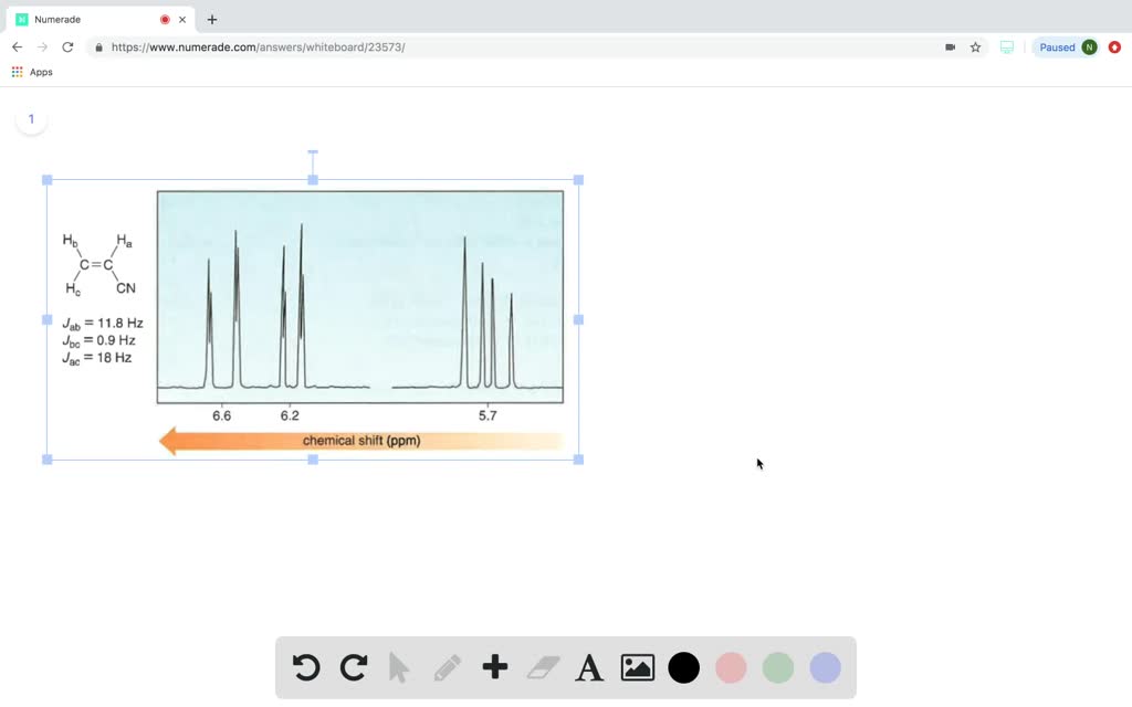
PDF ANALYSIS OF H NMR SPECTRA - University of Texas at Dallas spectrum to five signals, corresponding to 5 different sets of protons. 2. RELATIVE INTENSITIES OF THE PEAKS, OR SIGNALS. The term intensity, when used in reference to NMR signals, indicates the area under the peak. The areas under the peaks are given by an integrator when recording a spectrum.
A Step-By-Step Guide to 1D and 2D NMR Interpretation - Emery Pharma Now we have a fully assigned ¹H-NMR spectrum! This spectrum will help us assign our carbons using HSQC and HMBC NMR spectroscopy. Step 3: ¹³C-NMR Carbon NMR is a necessary step in full structural characterization. However, ¹³C-NMR alone does not provide enough information to assign the carbons in the molecule.
Publication Quality PDFs of NMR Spectra with TopSpin 3.5 and 3.6 Click the arrow next to Print and click Set Target Printer... under the sub-menu. Select the spectrum in the plot layout window. Check the box for Labels under Peaks . 9. Resize the spectrum so the red dotted line around the spectrum aligns with the gray dotted line representing the printable area of the page. 10.
Deciphering 1H NMR Spectra - StudyOrgo.com Once a NMR spectrograph is recorded, 4 pieces of information can be determined from the data as long as the chemical formula of the compound is known. To illustrate the points, we will consider the following 1H-NMR spectrum of the C 5 H 10 O. Signal Count - Number of unique hydrogens This is the easiest to interpret.
high resolution nuclear magnetic resonance (nmr) spectra - chemguide Look for NMR peaks in the 6.0 - 9.0 range. If you are given a number like 5 or 4 alongside that peak, this just tells you how many hydrogen atoms are attached to the ring. If there are 5 hydrogens attached to the ring, then there is only one group substituted into the ring.
6.7 ¹H NMR Spectra and Interpretation (Part II) With the structure of a compound given, we can apply all the knowledge about 1 H NMR to assign the signals in the spectrum to identify which signals come from which hydrogen (s). Examples Match the 1 H NMR spectrum below to its corresponding compound, and assign all of the signals. a) cyclopentanone b) 3-pentanone c) butaldehyde d) 2-pentanone
NMR Sample Preparation - Iowa State University WebLabel your sample. Permanent marker can be used to label the sample to ensure you do not mix up your NMR tube with another NMR user, or it can be retrieved at a later time. If you use a sticker or tape to label the tube, the sticker must be flush with the tube so it does not interfere with insert, eject or spinning inside the magnet.
Collaborative Computational Project for NMR - CCPN AnalysisScreen. Our latest AnalysisScreen developments make this a leading software tool for the analysis of NMR-based screening data: speed-optimised throughout, improved matching between reference and control spectra, flexible plotting functions and real-time updates when changing algorithms, thresholds or scoring functions.
PDF HOW TO PLOT AND PRINT NMR SPECTRUM - Swarthmore College FIRST, CLICK THE SIDE OF THE REPORT IMAGE WHERE THERE IS NO OBJECT. THE LEFT SIDE TOOLBAR WILL DISPLAY THE OPTION TO ADD A NMR SPECTRUM. USING THE MOUSE, LEFT CLICK-HOLD AND DRAG TO FORM A BOX WHERE THE INSET WILL BE. CLICKING INSIDE THE INSET SPECTRUM YOU CAN CHANGE THE SCALE, SIZE, X AND Y-AXIS LIMITS LIKE THE MAIN SPECTRUM.
Chapter 13: Nuclear Magnetic Resonance (NMR) Spectroscopy Webarbitrary label (your book uses X) 2.Ask what is the relationship of the compounds with the arbitrary label 3.If the labeled compounds are identical (or enantiomers), then the original nuclei are chemically equivalent and will normally give rise to a single resonance in the NMR spectra If the labeled compounds are not identical (and not ...
NMR Spectroscopy - Michigan State University The following features lead to the nmr phenomenon: 1. A spinning charge generates a magnetic field, as shown by the animation on the right. The resulting spin-magnet has a magnetic moment ( μ) proportional to the spin. 2. In the presence of an external magnetic field ( B0 ), two spin states exist, +1/2 and -1/2.
How To Label NMR Spectra - YouTube About Press Copyright Contact us Creators Advertise Developers Terms Privacy Policy & Safety How YouTube works Test new features Press Copyright Contact us Creators ...
How to Interpret Proton NMR Spectra - theSpectroscopy Now that we have had an introduction to key aspects of 1 H NMR spectra (chemical shift, peak area, and signal splitting), we can start to apply 1 H NMR spectroscopy to elucidating the structure of unknown compounds. The following steps summarize the process:
How to interpret NMR spectra - University of Texas at Austin 1.2 An NMR spectrum is a plot of absorbance versus frequency. 1.2A To make different spectra directly comparable, a standard is used for all NMR spectra. For 1H NMR spectra, the standard is called tetramethylsilane (TMS) and a small amount of TMS is usually added to any 1H NMR sample.
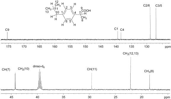
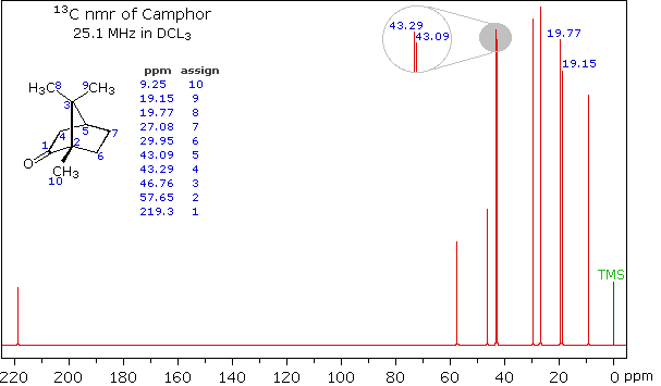
interpreting C-13 NMR spectra - chemguide Using the simplified table of chemical shifts above, work out the structure of the compound with the following C-13 NMR spectrum. Its molecular formula is C 4 H 6 O 2. Let's sort out what we've got. There are four peaks and four carbons. No two carbons are in exactly the same environment.
MetaboAnalyst WebGroup label - 0, 15, 30, ... Binned 1H NMR spectra of 50 urine samples using 0.04 ppm constant width (Psihogios NG, et al.) Group 1- control; group 2 - severe kidney ... Compound concentrations of 14 urine samples collected from 7 cows at two time points using 1H NMR (unpublished data). Group 1- day 1, group 2- day 4 ...
NMR Spectrum of Phenacetin | Thermo Fisher Scientific - US Phenacetin (C 10 H 13 NO 2 ), N- (4-Ethoxyphenyl)acetamide, is a drug that saw widespread application for nearly a century in many over-the-counter medications as a remedy for pain and fever. The 82 MHz spectrum of a 6% (w/w; 0.5 M) solution of phenacetin in CDCl 3 acquired using the Thermo Scientific picoSpin 80 NMR spectrometer.
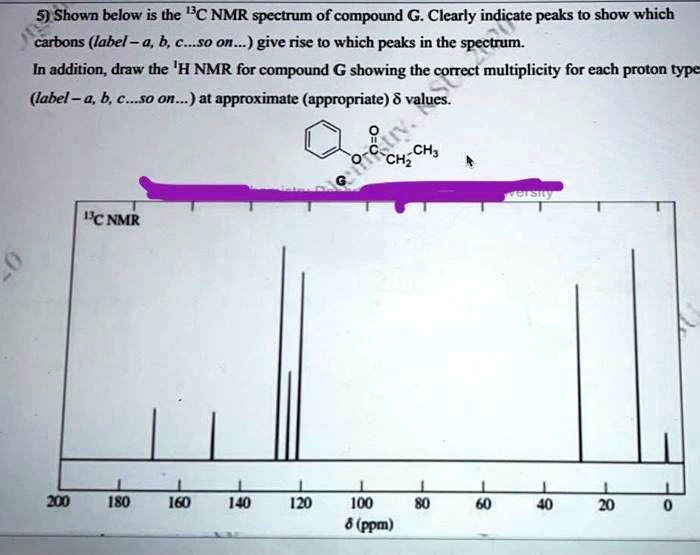
PDF Examination of Proton NMR Spectra considerably easier than analysis of proton spectra. In most cases the analysis of a C-13 spectrum involves noting the number of signals and their chemical shift. Each carbon gives rise to a signal, but if two or more carbons are equivalent, their signals will exactly overlap. As in proton NMR chemical shift may suggest the type of
Interpreting a C-13 NMR spectrum - Vassar College DEPT-135 Spectrum. The chemical shifts of the peaks on these two spectra are the same as they are on the C-NMR spectrum. The sizes of the peaks on the two spectra are different, and this size difference is the key to understanding them. You may have noticed the peak at 35 ppm on the C-NMR spectrum doesn't appear on either of the DEPT spectra ...
NMR Spectroscopy | ChemTalk C-13 NMR spectroscopy is specific to the analysis of the carbons in a molecule. It is less common than H-1 but is still regularly used in organic chemistry laboratories. C-13 NMR can give the following information: The number of unique carbons in the compound. Chemical environment of the compound.
NMR Spectroscopy Principles, Interpreting an NMR Spectrum and Common ... By convention, the chemical shift scale in an NMR spectrum is represented from right to left. As described above, the zero value is established using a standard compound whose carbon and hydrogen atoms are strongly shielded and hence, their signals appear in the furthest upfield region (as seen in Figure 7).
How to Identify Molecular Fragments from NMR and IR Spectra The IR and NMR spectra for a compound with molecular formula C 5 H 10 O Carbonyl groups are found in aldehydes, ketones, esters, and carboxylic acids. Here, however, esters and carboxylic acids can be eliminated because the molecular formula shows only a single oxygen.
MetaboAnalyst Group Label. Description: Data 1 ... Common name: Discrete: Urinary metabolite concentrations from 77 cancer patients measured by 1H NMR. ... You can still process ...
Introduction to CREST — xtb doc 2021 documentation - Read the … WebOther standalone functionalities that are included in CREST are parallel optimization and screening functions for GFNn–xTB, the function to sort (e.g. for NMR equivalencies) externally created ensembles, and some automated procedures for the protonation, deprotonation and tautomerization of structures.
Starting guide to Mnova 1D-NMR spectrum - Mestrelab Resources To do this go to 'NMR/Processing' and then apply 'Auto baseline correction'. This spectrum is now ready for analysis so go now to 'NMR/Analysis' and select 'Reference' to make sure that this peak is set to the suitable chemical shift. Now, click 'Auto Multiplet Analysis' button to carry out Peak picking, Integration and ...














Post a Comment for "39 how to label nmr spectra"