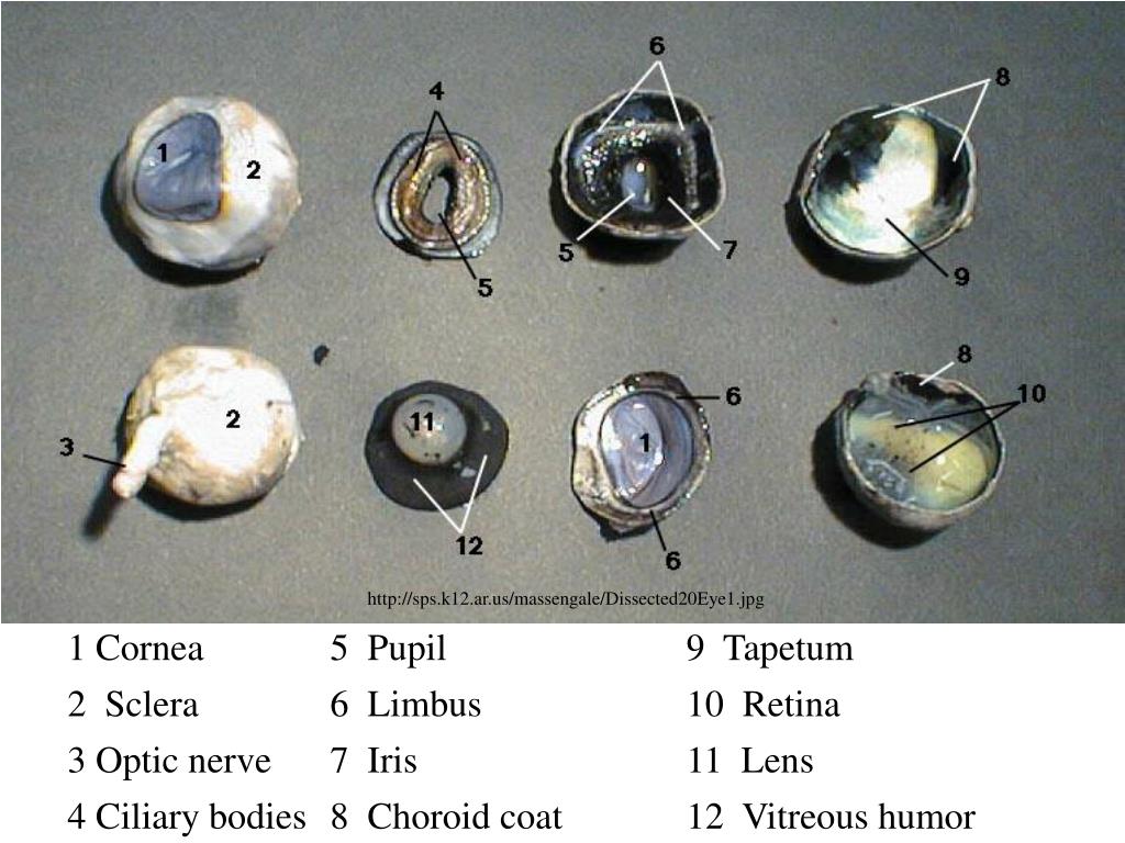
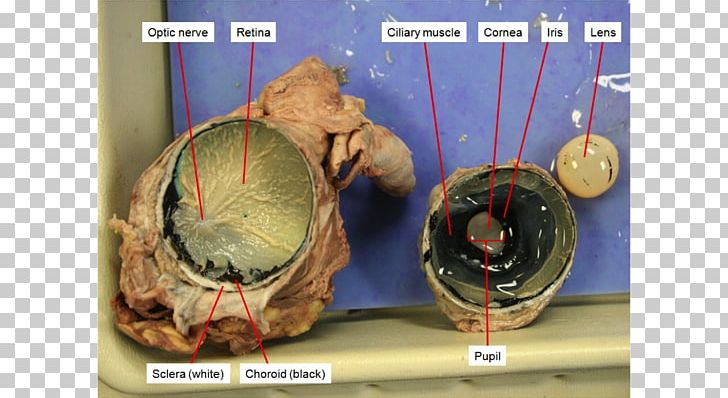
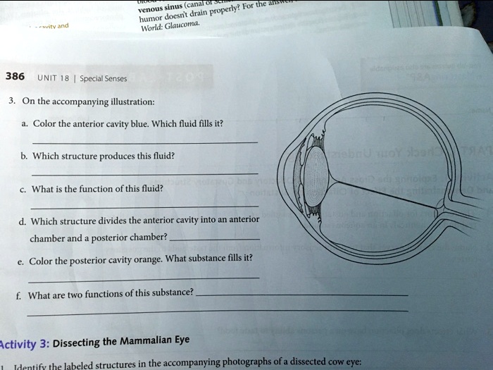
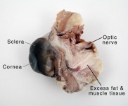
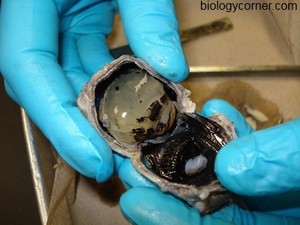
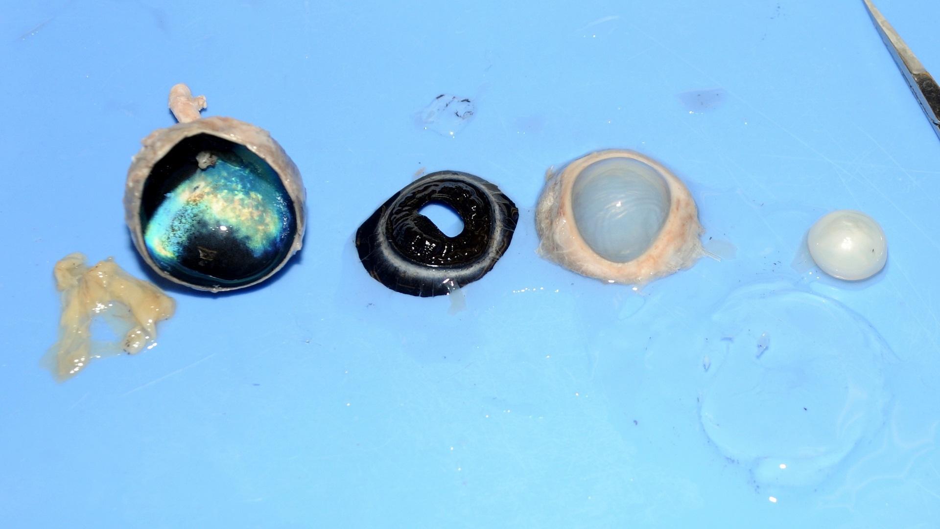
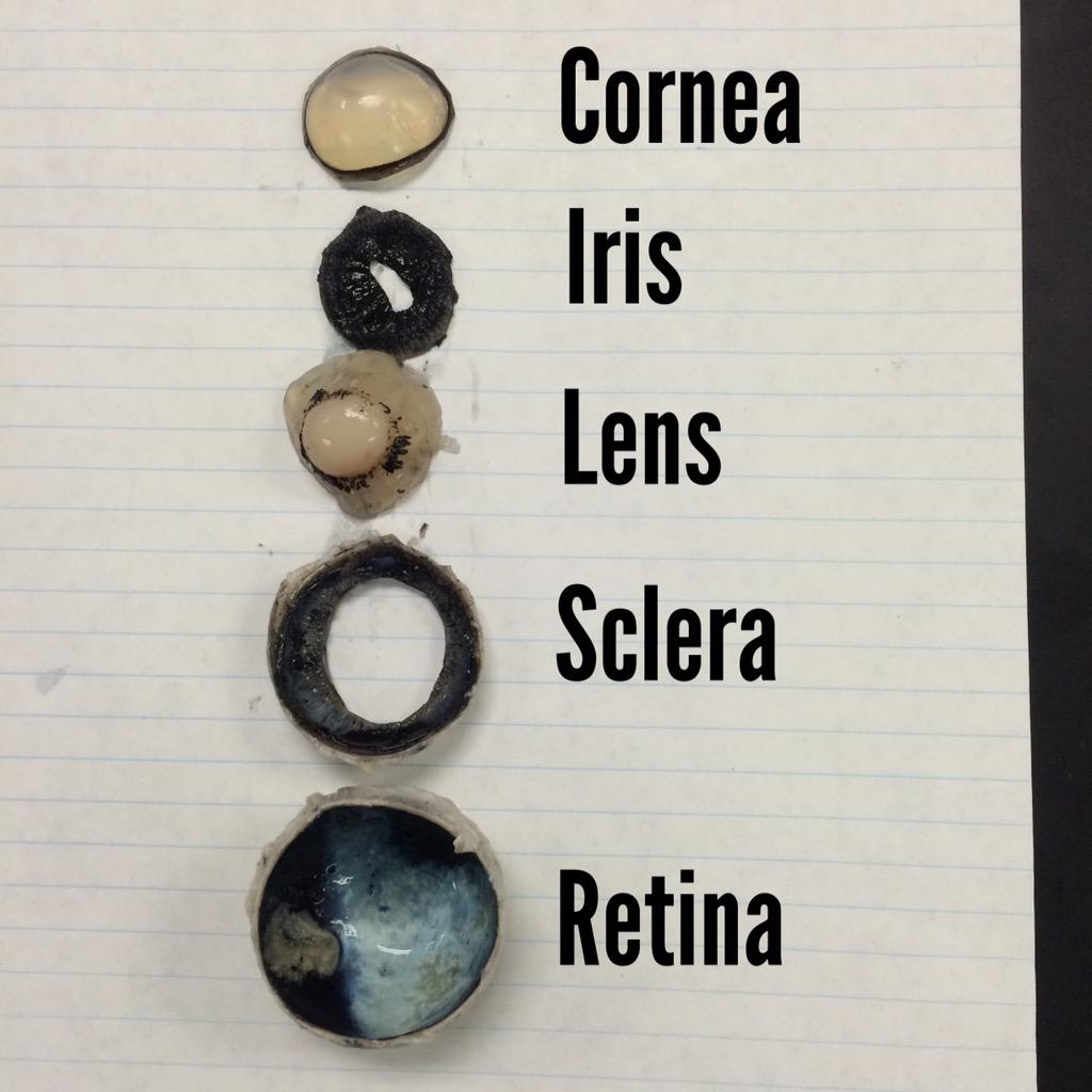
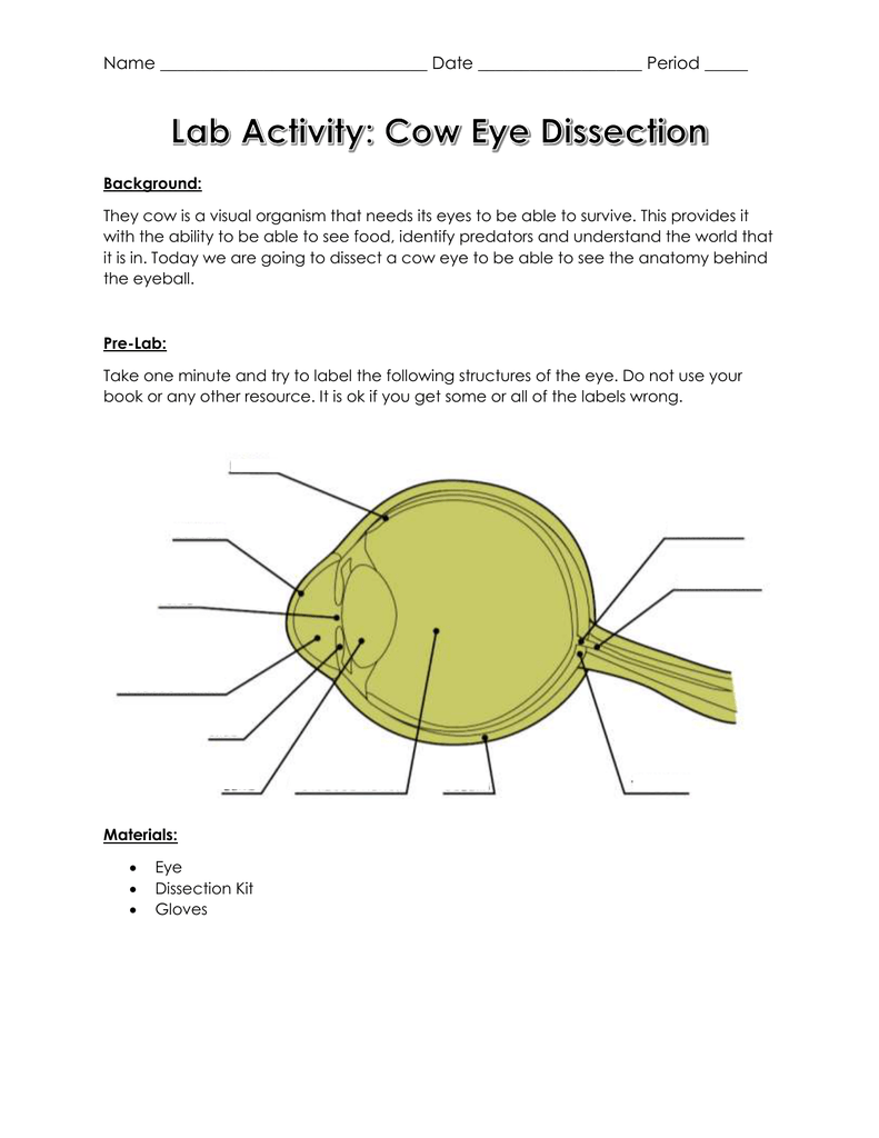
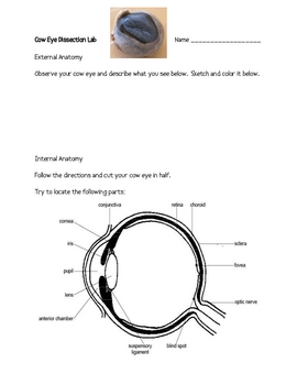
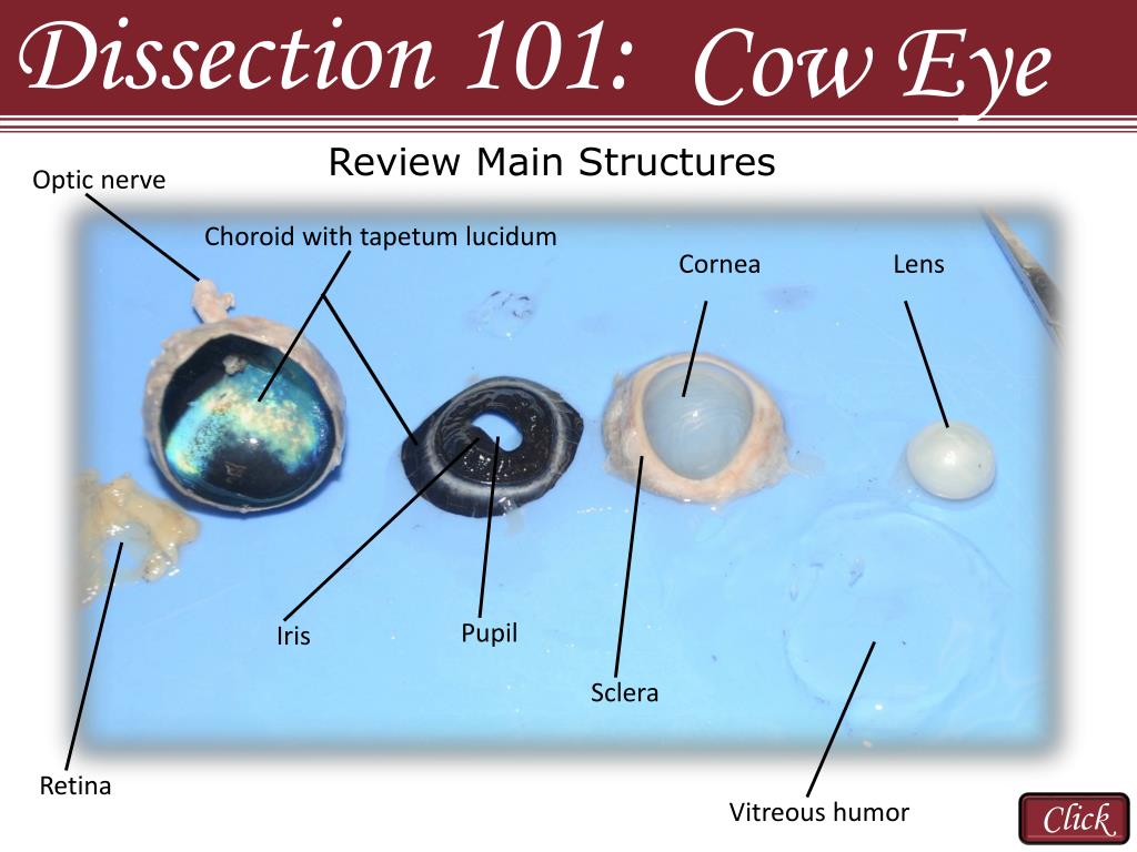
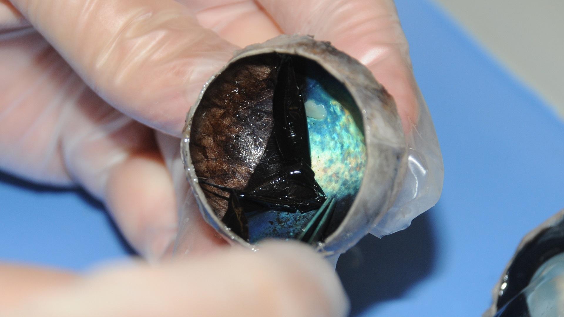
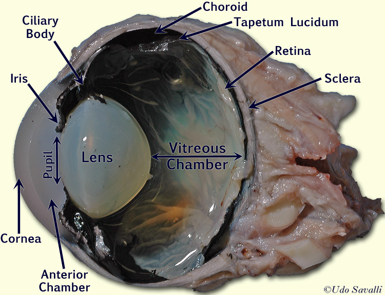
38 dissected cow eye labeled
Anatomy Lab Preserved Cow Eye Specimen for Dissection, for Biology ... Cow Eye Dissection: Anatomy Lab's Preserved Cow Eye specimens are perfect for studying human and other mammalian eye anatomy. It's an economical way to study the mammalian organ's structure and details, including the cornea, lens, retina and iris. The large size of the eye aids in the identification of the internal anatomy of the eye. Cow Eye Dissection - The Biology Corner COW EYE DISSECTION 1. Examine the outside of the eye. You should be able to find the sclera, or the whites of the eye. This tough, outer covering of the eyeball has fat and muscle attached to it 2. Locate the covering over the front of the eye, the cornea. When the cow was alive, the cornea was clear.
Cow Eye Dissection Guide - Google Slides Dissection 101: Cow Eye Use the point of a scissors or a scalpel to make an incision through the layers of the eye capsule (similar to figure 1); there are three layers from the exterior:...
Dissected cow eye labeled
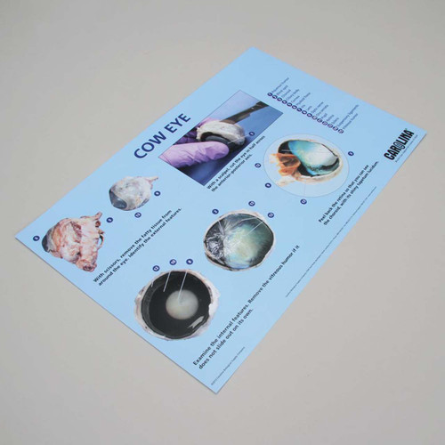
Cow eye - dissection and label Cow eye shown with labeled cornea. The cornea is the transparent front part of the eye that covers the iris, pupil, and anterior chamber. The cornea, with the anterior chamber and lens, refracts light, with the cornea accounting for approximately two-thirds of the eye's total optical power. 3. Lab: Cow Eye Dissection Flashcards | Quizlet Anatomy Lab Practical #4 (Cow Eye Dissec… 12 terms. sdaly724. Cow Eye Dissection. 15 terms. Ma_Petite_Ballerina. Tool Id. 17 terms. Brywar99. Lab: Cow Eye Dissection. 14 terms. enthusiastic_crafter. Recent flashcard sets. ACT 2. 8 terms. Lauren_Ogrodowicz. bio 2 final exam. 26 terms. Cow Eye Dissection - Carolina Knowledge Center Carolina's Perfect Solution® cow eye dissection introduces students to the anatomy of the mammalian eye. This activity allows students to identify the major structures of the eye. The activity supports 3-dimensional learning and builds toward the following: NGSS Scientific and Engineering Practice: Developing and Using Models
Dissected cow eye labeled. Steinhagen, North Rhine-Westphalia - Wikipedia Steinhagen, North Rhine-Westphalia. An Steinhagen amo in usa ka munisipalidad ha distrito han Gütersloh, ha North Rhine-Westphalia, Alemanya . k. h. l. Usa ka turók ini nga barasahon. Dako it imo maibubulig ha Wikipedia pinaagi han pagparabong hini. An Wikimedia Commons mayda media nga nahahanungod han: Steinhagen, North Rhine-Westphalia. Cow Eye Dissection | Carolina.com Cow Eye Internal Anatomy Hold the eye between your thumb and forefinger, as shown below. Using scissors or a scalpel, carefully cut the eye in half, separating the front and back of the eye. Examine the inside front portion of the eye. Remove the gelatinous vitreous humor and hard lens. Cow Eye Dissection Lab from Anatomy and Physiology Cow Eye Dissection Lab from Anatomy and Physiology These labs were assigned in A&P I and II University Community College System of New Hampshire Course Nursing (ADNR 116) Academic year2020/2021 Helpful? 163 Comments Please sign inor registerto post comments. Students also viewed shock practice questions Chapter 9 - Practice Assignment Cow Eye Dissection - Perkins School for the Blind Cow Eye Dissection Step-by-step instructions for a science lab to dissect a cow eye SHARE This is a great activity for middle and high school students. It allows them to participate in a dissection, while teaching the anatomy and structure of the eye. To get started, watch the YouTube video Cow's Eye Dissection: Exploratorium.
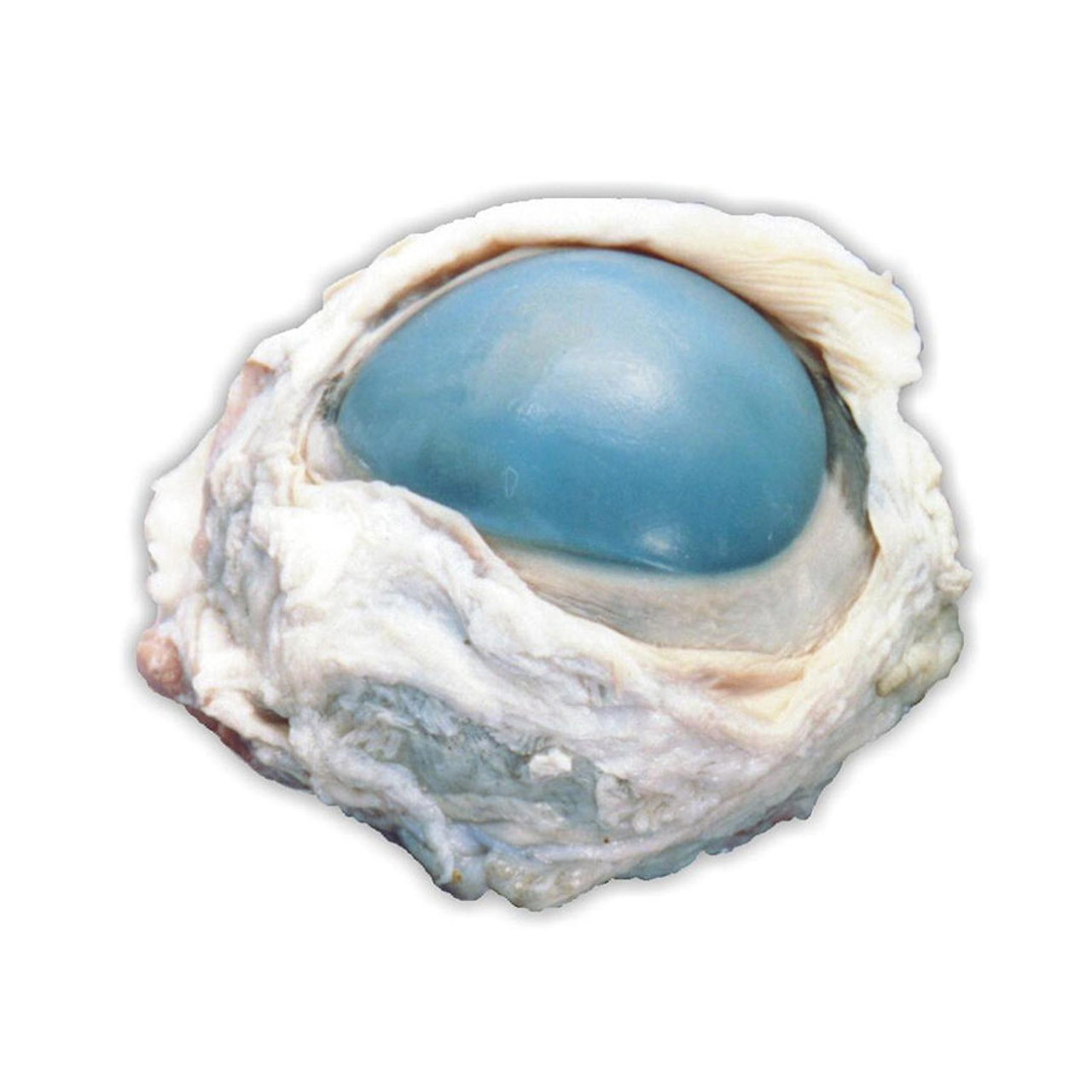
Preserved Cow Eye Dissection Specimen | Anatomy Lab Preserved Specimen: Cow Eye. Anatomy Lab's Preserved Cow Eye Specimen is perfect for learning about the internal and external structures of the organ. Learn comparative anatomy with this specimen by comparing its structures to other mammals, including humans. This dissection specimen is also great for understanding the physiology and function ... anatomy and physiology of cow - Microsoft anatomy sheep dissection heart side dissected pig physiology science human class card labeled medical septum cow notes parts interventricular study. Pin On Anatomy And Physiology . eye worksheet diagram human cow dissection anatomy worksheets structure physiology answers eyeball drawing label body labeling eyes ear template muscle 13.7: Cow Eye Dissection - Biology LibreTexts 1. Examine the outside of the eye. You should be able to find the sclera, or the whites of the eye. This tough, outer covering of the eyeball has fat and muscle attached to it 2. Locate the covering over the front of the eye, the cornea. When the cow was alive, the cornea was clear. In your cow's eye, the cornea may be cloudy or blue in color. 2. Cow Eye Dissection & Anatomy Project | HST Learning Center Place the cow's eye on a dissecting tray. The eye most likely has a thick covering of fat and muscle tissue. Carefully cut away the fat and the muscle. As you get closer to the actual eyeball, you may notice muscles that are attached directly to the sclera and along the optic nerve.
Cow Eye Labeling Quiz - PurposeGames.com This online quiz is called Cow Eye Labeling. It was created by member secretsquirrel and has 11 questions. ... Label Lateral View Of The Brain. Science. English. Creator. EllenEllen. Quiz Type. Image Quiz. Value. 10 points. Likes. 17. Played. 56,880 times. Printable Worksheet. Play Now. Add to playlist. Cow Eye Dissection - YouTube Cow Eye Dissection - YouTube 0:00 / 15:03 Cow Eye Dissection Biologybyme 23.8K subscribers Subscribe 13K 1.5M views 9 years ago Dissections - Biologybyme Show more Heart dissection (Pig)... Cow Eye Dissection & Parts of the Eye Diagram | Quizlet Clear, outer layer of the front of the eye. sclera White, outermost layer of the eye. Helps maintain shape and gives attachment to muscles. photoreceptors The cells in the retina that respond to light (rods and cones) rods Photoreceptor cells in the eye that detect black, white, and gray cones Photoreceptor cells in the eye that detect color Solved 1. Identify the labeled structures in the | Chegg.com Identify the labeled structures in the accompanying photographs of a dissected cow eye. a. Sclera b. Cornea c. choroid? d. Optic Disc - Retina UNIT 17 | General and Special Senses 395 Show transcribed image text Expert Answer 1st step All steps Answer only Step 1/5 Label a Sclera
PDF COW'S EYE dissection - Exploratorium COW'S EYE dissection page 6 Now take a look at the rest of the eye. If the vitreous humor is still in the eyeball, empty it out. On the inside of the back half of the eyeball, you can see some blood vessels that are part of a thin fleshy film. That film is the retina. Before you cut the eye open, the vitreous humor
Animal Organ Dissection Kit | Mammalian Anatomy Lab for Kids Mammal Organs Dissection Kit $51.95 Study animal mammalian organ anatomy as you dissect the four preserved specimens in this kit: a cow eye, sheep heart, sheep brain & sheep kidney. Includes dissecting tools & dissection guides for all four specimens! Out of Stock, Expected to Ship: 3/14/2023 Ages 11+ Shipping Restrictions share Add to Wish List
Cow Eye Dissection Quiz - PurposeGames.com Cow Eye Dissection by avantitreasures787 67,383 plays 8 questions ~20 sec English 8p More 33 too few (you: not rated) Tries Unlimited [?] Last Played February 22, 2022 - 12:00 am From the quiz author cow eye, dissection Remaining 0 Correct 0 Wrong 0 Press play! 0% 0:00.0 Show More Other Games of Interest Bones of the foot and ankle Science English
Nerous System Lab 08 - ©eScience Labs, 2016 The Nervous ... - StuDocu the nervous system experiment cow eye dissection questions how does the eye work? the eye works like camera. light enters the eye through cornea, the cornea. Skip to document. Ask an Expert. ... Anatomy & Physiology I 80% (5) 1. COW EYE LAB Week 8 dissection post lab questions. Anatomy & Physiology I 80% (5) English (US) United States. Company ...
Dissecting An Eyeball - Krieger Science Returning to our dissection, underneath the retina is a pretty, shiny, blue-green mirror, officially called the tapetum. This is what causes cow's eyes to shine in headlights, and it is probably the most memorable part of an eye dissection for children. This is also one thing that cows and people do not have in common.
Cow Eye Dissection Kit for Kids Animal Anatomy Labs | HST Cow Eye Dissection Kit $11.30 This Cow Eye Dissection Kit gives an inside view of how the eye works. It comes with everything you need for this activity, including a preserved cow eye specimen, a step-by-step dissection guide & essential dissection tools. quantity Ages 11+ In Stock & Ready to Ship Need it fast? See delivery options in cart.
13.7: Cow Eye Dissection - Medicine LibreTexts 13.7: Cow Eye Dissection. 1. Examine the outside of the eye. You should be able to find the sclera, or the whites of the eye. This tough, outer covering of the eyeball has fat and muscle attached to it. 2. Locate the covering over the front of the eye, the cornea. When the cow was alive, the cornea was clear.
Eye Dissection - BIOLOGY JUNCTION By dissecting the eye of a cow, which is similar to the eyes of all mammals including humans, you will gain an understanding of the structure and function of the parts of the eye. Materials: Cow eye, dissecting pan, dissecting kit, safety glasses, lab apron, and gloves Procedure (External Structure):
Solved PART C: Assessments Complete the following: FIGURE - Chegg Anatomy and Physiology. Anatomy and Physiology questions and answers. PART C: Assessments Complete the following: FIGURE 35.14 Partial frontal cut of dissected cow eye. Label the internal structures using the list provided. 3 Ciliary body Lens Retina Sclera Tapetum fibrosum of choroid) Question: PART C: Assessments Complete the following ...
Cow's Eye Dissection - Eye diagram - Exploratorium A cow's iris is brown. Human irises come in many colors, including brown, blue, green, and gray. A clear fluid that helps the cornea keep its rounded shape. The pupil is the dark circle in the center of your iris. It's a hole that lets light into the inner eye. Your pupil is round. A cow's pupil is oval.
PDF Home Science Tools Resource Center Home Science Tools Resource Center
Cow Eye Dissection - Carolina Knowledge Center Carolina's Perfect Solution® cow eye dissection introduces students to the anatomy of the mammalian eye. This activity allows students to identify the major structures of the eye. The activity supports 3-dimensional learning and builds toward the following: NGSS Scientific and Engineering Practice: Developing and Using Models
Lab: Cow Eye Dissection Flashcards | Quizlet Anatomy Lab Practical #4 (Cow Eye Dissec… 12 terms. sdaly724. Cow Eye Dissection. 15 terms. Ma_Petite_Ballerina. Tool Id. 17 terms. Brywar99. Lab: Cow Eye Dissection. 14 terms. enthusiastic_crafter. Recent flashcard sets. ACT 2. 8 terms. Lauren_Ogrodowicz. bio 2 final exam. 26 terms.
Cow eye - dissection and label Cow eye shown with labeled cornea. The cornea is the transparent front part of the eye that covers the iris, pupil, and anterior chamber. The cornea, with the anterior chamber and lens, refracts light, with the cornea accounting for approximately two-thirds of the eye's total optical power. 3.














Post a Comment for "38 dissected cow eye labeled"